
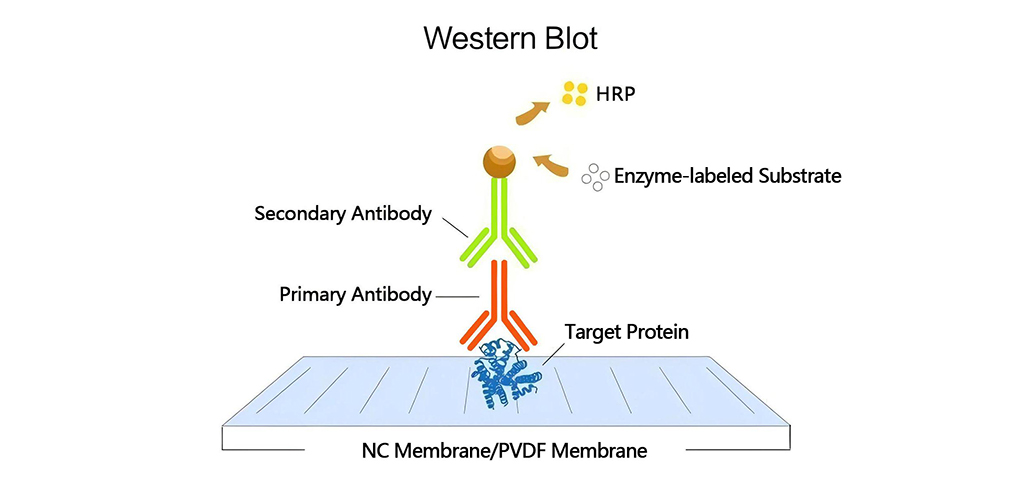
Western Blotting (WB), also known as protein immunoblotting, is a technique that transfers total proteins from cells or tissues via electrophoresis from a gel to a solid support membrane (such as nitrocellulose membrane NC or polyvinylidene fluoride membrane PVDF), followed by detection of specific antigens using antibodies. This technology is widely used in protein expression studies, antibody activity detection, and disease diagnosis.
PVDF Membrane Usage Guide
-Pretreatment Steps
PVDF membranes are hydrophobic and require the following pretreatment:
- Wet Transfer Method:
- Soak in methanol for 30 seconds (membrane changes from opaque to semi-transparent)
- Soak in double-distilled water for 2 minutes
- Equilibrate in transfer buffer for ≥5 minutes
- Semi-Dry Transfer Method:
- Soak in methanol for 15 seconds
- Subsequent steps are the same as the wet transfer method
-Pore Size Selection
Pore Size | Applicable Molecular Weight | Characteristics | Application Scenarios |
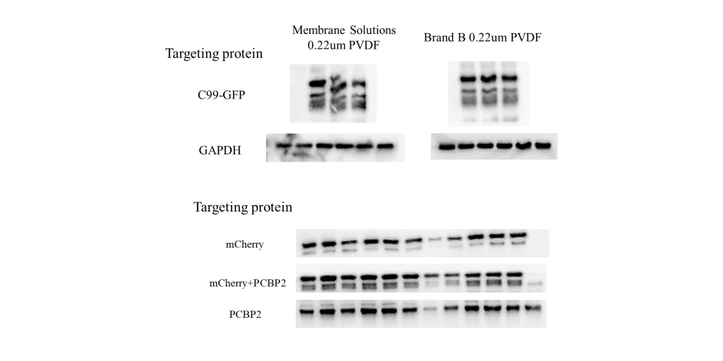
0.22μm | <20kD | High binding capacity, high resolution | Small protein detection, precise quantification |
0.45μm | >20kD | High throughput, low operational resistance | Large protein and nucleic acid transfer |
Transfer Efficiency Verification
- Reversible Staining (Recommended):
- Common dye: 0.2% Ponceau S (diluted in 1% acetic acid)
- Steps: Wet with methanol → Stain for 1 minute → Rinse with double-distilled water → Completely remove with 0.1N NaOH
- Irreversible Staining:
- Dyes: Amido Black, Coomassie Brilliant Blue
- Characteristics: High sensitivity but interferes with subsequent analysis
-Membrane Selection: PVDF vs NC
Property | PVDF Membrane | NC Membrane |
Physical Properties | High strength, good flexibility | Brittle, prone to breakage |
Chemical Stability | Resistant to acids, bases, and organic solvents | Sensitive to acids and bases |
Temperature Range | -10°C~150°C | -10°C~40°C |
Transparency | High | Milky white |
Cost | Higher | Lower |
Applications | Harsh conditions, small proteins, multiple detections, long-term storage | Routine detection, large proteins, fluorescent labeling, low-budget projects |
Key Considerations
-Transfer Structure Preparation:
- Thoroughly clean transfer equipment
- Carefully remove bubbles in the “sandwich” structure
- Ensure tight contact between gel and membrane
-Transfer Condition Optimization:
- Maintain temperature control with ice bath
- Standard conditions: 1.0mm thick 10% gel, 300mA transfer for 1-1.5 hours (250kD protein)
- Small proteins (<20kD):
- Omit SDS from transfer buffer
- Add 10-20% methanol
- Use 0.22μm PVDF membrane
- Reduce transfer intensity (200-250mA, 15-30min)
-Antibody Stripping and Membrane Storage:
- Stripping: Use a combination of heating and detergents
- Storage:
- Short-term: 4°C (≤2 weeks)
- Medium-term: -20°C (≤2 months)
- Long-term: -70°C
- Storage method: Sandwich between filter paper and cardstock, store in sealed bags
Technical Summary
- Prioritize 0.22μm PVDF membranes for small protein detection
- Temperature control during transfer is critical
- Ponceau S staining is the preferred method for verifying transfer efficiency
- PVDF membranes are suitable for harsh experimental conditions and long-term storage
- NC membranes are better for routine detection and cost-sensitive projects
By strictly adhering to the above operational standards and technical points, reliable and highly reproducible Western Blotting results can be achieved.


Leave a Comment
Your email address will not be published. Required fields are marked *