U-Blot® OPA1 Rabbit mAb
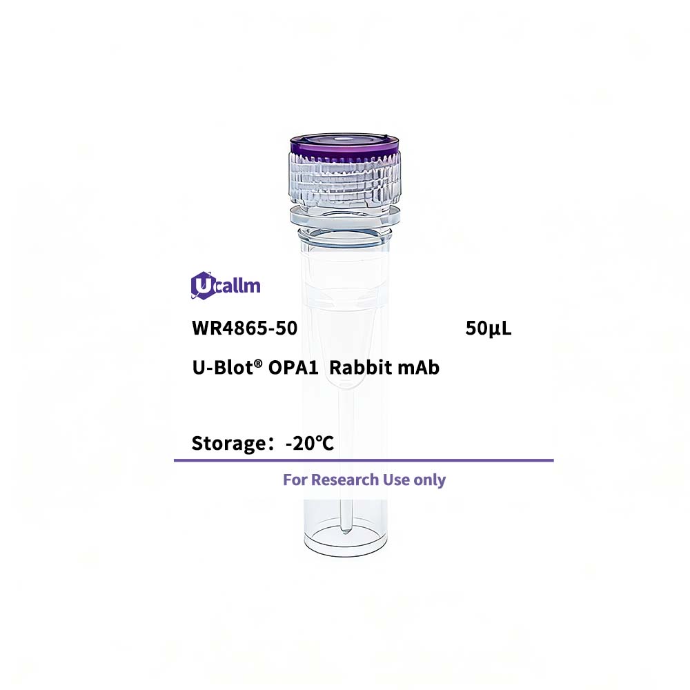
Ucallm_WR4865-50
Price range: $268.00 through $328.00
Product details
Background:This gene product is a nuclear-encoded mitochondrial protein with similarity to dynamin-related GTPases. It is a component of the mitochondrial network. Mutations in this gene have been associated with optic atrophy type 1, which is a dominantly inherited optic neuropathy resulting in progressive loss of visual acuity, leading in many cases to legal blindness. Multiple transcript variants encoding different isoforms have been found for this gene. [provided by RefSeq, Mar 2009],
Specifications
| Size | 50μL/100μL |
| Target | OPA1 |
| Reactivity | Human, Mouse, Rat |
| Applications | WB, IHC, IF, ELISA |
| MW(Calculated) | 112kD |
| MW(Observed) | 80kD |
| Host Species | Rabbit |
| Isotype | IgG,Kappa |
| Conjugate/Modification | Unmodified |
| Recommended Dilution Ratio | IHC 1:1000-1:4000; WB 1:10000-1:50000; IF 1:200-1:1000; ELISA 1:5000-1:20000; / Note: For IHC, we suggest antigen retrieval with TE buffer pH 9.0 (Cat#WT1004) |
| Formulation | PBS, 50% glycerol, 0.05% Proclin 300, 0.05%BSA |
| Purification | Recombinant Antibody expressed in animal component-free (ACF) media, purified via Protein A affinity chromatography. |
| Storage | -15°C to -25°C/1 year(Do not lower than -25°C) |
| Concentration | |
| Clonality | Monoclonal |
| Clone Number | RM1035R |
| Immunogen | The specific immunogen used to produce this antibody is proprietary information. |
| Specificity | Endogenous |
| Gene Name | OPA1 KIAA0567 |
| Protein Name | Dynamin-like 120 kDa protein, mitochondrial (Optic atrophy protein 1) |
| Other Name | |
| Species | Human |
| Gene ID | 4976 |
| Uniprot | O60313, |
| Species.1 | Mouse |
| Gene ID.1 | |
| Uniprot.1 | P58281, |
| Species.2 | Rat |
| Gene ID.2 | |
| Uniprot.2 | Q2TA68 |
| Cellular Localization | Mitochondrion inner membrane; Single-pass membrane protein . Mitochondrion intermembrane space . Mitochondrion membrane . Detected at contact sites between endoplasmic reticulum and mitochondrion membranes. . |
| Tissue Expression | Highly expressed in retina. Also expressed in brain, testis, heart and skeletal muscle. Isoform 1 expressed in retina, skeletal muscle, heart, lung, ovary, colon, thyroid gland, leukocytes and fetal brain. Isoform 2 expressed in colon, liver, kidney, thyroid gland and leukocytes. Low levels of all isoforms expressed in a variety of tissues. |
| Signaling_pathway | Cellular Processes >> Transport and catabolism >> Mitophagy - animal;Human Diseases >> Neurodegenerative disease >> Spinocerebellar ataxia |
| Research Areas | >>Spinocerebellar ataxia |
| Function | Disease:Defects in OPA1 are a cause of optic atrophy type 1 (OPA1) [MIM:165500]. OPA1 is a dominantly inherited optic neuropathy occurring in 1 in 50,000 individuals that features progressive loss in visual acuity leading, in many cases, to legal blindness.,Disease:Defects in OPA1 are the cause of optic atrophy 1 and deafness [MIM:125250]. Some individuals with mutations in OPA1 manifest also ophthalmoplegia and myopathy.,Function:Dynamin-related GTPase required for mitochondrial fusion and regulation of apoptosis. May form a diffusion barrier for proteins stored in mitochondrial cristae. Proteolytic processing in response to intrinsic apoptotic signals may lead to disassembly of OPA1 oligomers and release of the caspase activator cytochrome C (CYCS) into the mitochondrial intermembrane space.,PTM:PARL-dependent proteolytic processing releases an antiapoptotic soluble form not required for mitochondrial fusion.,similarity:Belongs to the dynamin family.,subunit:Oligomeric complex consisting of membrane-bound and soluble forms of OPA1. Binds PARL.,tissue specificity:Highly expressed in retina. Also expressed in brain, testis, heart and skeletal muscle. Isoform 1 expressed in retina, skeletal muscle, heart, lung, ovary, colon, thyroid gland, leukocytes and fetal brain. Isoform 2 expressed in colon, liver, kidney, thyroid gland and leukocytes. Low levels of all isoforms expressed in a variety of tissues., |
Please login to post questions
Related products