
Introduction
Western Blot (WB) is a widely used technique in molecular biology, biochemistry, and medical research for detecting and quantifying specific proteins in a complex mixture. By combining gel electrophoresis with immunoblotting, Western Blot allows researchers to analyze protein expression, verify protein modifications, and assess molecular weight with high specificity and sensitivity. This article provides an in-depth look at the principles, workflow, and applications of Western Blot, highlighting its importance in scientific and clinical research.
Principles of Western Blot
The Western Blot technique relies on three fundamental steps:
1.Protein Separation– Proteins are separated based on their molecular weight using sodium dodecyl sulfate-polyacrylamide gel electrophoresis (SDS-PAGE).
2.Protein Transfer– Separated proteins are transferred onto a solid support membrane (PVDF or nitrocellulose) for analysis.
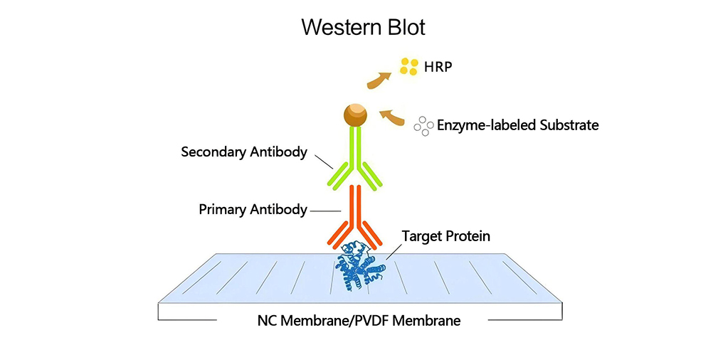
3.Antibody-Based Detection– Specific proteins are identified using primary and secondary antibodies conjugated to detection systems such as chemiluminescence or fluorescence.
Workflow of Western Blot
1. Sample Preparation
- Proteins are extracted from cells or tissues using a lysis buffer containing protease and phosphatase inhibitors.
- Protein concentration is quantified to ensure equal loading.
- The sample is denatured using heat and SDS to provide uniform charge distribution.
2. Gel Electrophoresis
- Proteins are loaded into a polyacrylamide gel and separated based on size under an electric field.
- A molecular weight marker (protein ladder) is included to estimate protein sizes.
3. Protein Transfer
- Separated proteins are transferred from the gel to a membrane (PVDF or nitrocellulose) via electroblotting.
- Ponceau S staining can be used to verify the successful transfer.
4. Blocking and Antibody Incubation
- The membrane is blocked with a solution (e.g., BSA or non-fat milk) to prevent non-specific binding.
- Primary antibodies specific to the target protein are incubated with the membrane.
- Secondary antibodies conjugated with HRP or fluorescent labels bind to the primary antibody for detection.
5. Detection and Analysis
- The membrane is treated with a substrate (e.g., enhanced chemiluminescence, ECL) to produce a detectable signal.
- The signal is visualized using imaging systems like X-ray films or digital CCD cameras.
- Band intensity is quantified for protein expression analysis using densitometry software.
Applications of Western Blot
Western Blot has extensive applications across various scientific fields:
1. Biomedical Research
- Detecting biomarkers for diseases such as cancer and neurodegenerative disorders.
- Studying protein-protein interactions and signaling pathways.
- Verifying gene expression at the protein level.
2. Clinical Diagnostics
- Diagnosis of infectious diseases (e.g., HIV, Lyme disease) by detecting pathogen-specific proteins.
- Confirmation of autoimmune diseases by identifying abnormal antibody-protein interactions.
- Quality control testing of pharmaceutical products.
3. Biopharmaceutical and Drug Development
- Evaluating the effects of drugs on protein expression and function.
- Assessing protein modifications (e.g., phosphorylation, glycosylation) relevant to therapeutic targets.
- Validating recombinant protein production in biotechnology.
Advantages and Limitations
1.Advantages
- High specificity due to antibody-based detection.
- Ability to quantify protein expression changes.
- Compatibility with a wide range of sample types (cell lysates, tissue extracts, serum, etc.).
2.Limitations
- Requires careful optimization of antibodies and experimental conditions.
- Time-consuming, involving multiple steps.
- Limited dynamic range compared to mass spectrometry-based proteomics.
Conclusion
Western Blot remains a cornerstone technique in protein research, offering precise detection and quantification capabilities for various applications. Whether in fundamental research, clinical diagnostics, or drug development, this powerful method continues to provide invaluable insights into protein biology and disease mechanisms. As advancements in imaging technology and antibody development continue, Western Blot will remain a critical tool in the life sciences for years to come.


Leave a Comment
Your email address will not be published. Required fields are marked *