
The viewpoint that phosphorylation fundamentally transforms cellular processes through precise molecular modifications is absolutely correct. As a matter of fact, this post-translational modification represents one of the most critical regulatory mechanisms in biological systems. This is mainly reflected in the following aspects of cellular control and signal transduction.
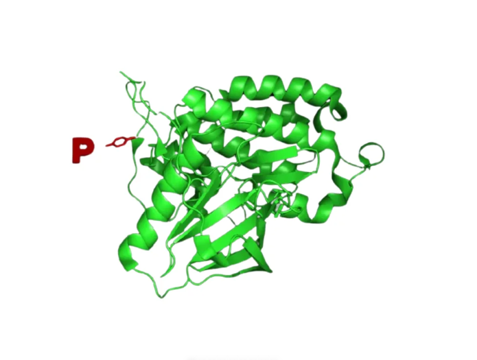
We observe that protein phosphorylation mechanism operates through kinase-catalyzed addition of phosphate groups to specific amino acid residues. These negatively charged modifications create conformational changes that dramatically alter molecular behavior. The process directly influences approximately 13,000 human proteins at their designated phosphorylation sites.
You will discover that this reversible modification serves as a molecular switch. Kinases transfer phosphate groups from ATP to serine, threonine, and tyrosine residues. The resulting structural changes activate or deactivate enzymatic activity, modify binding affinities, and alter cellular localization patterns.
Key Takeaways
- Phosphorylation creates reversible conformational changes that regulate cellular processes
- Protein kinases catalyze phosphate group transfer from ATP to specific amino acid residues
- Approximately 13,000 human proteins contain phosphorylation sites for regulatory control
- This modification serves as a molecular switch for activation and deactivation
- Phosphorylation influences enzyme activity, binding affinities, and protein localization
- The process primarily targets serine, threonine, and tyrosine residues
Introduction to Phosphorylation
Understanding phosphorylation requires exploring how cells use this dynamic modification to control protein activity and cellular responses. This biochemical process stands as one of the most important post-translational modifications in cellular biology. We observe phosphorylation occurring across all living organisms, from simple bacteria to complex human cells.
The reversible nature of phosphorylation makes it an ideal regulatory mechanism. Cells can rapidly activate or deactivate proteins based on immediate needs. This flexibility allows organisms to respond quickly to environmental changes and metabolic demands.
What is Phosphorylation?
Phosphorylation involves the enzymatic addition of phosphate groups to specific amino acid residues on proteins. This process specifically targets amino acids containing hydroxyl groups. In eukaryotic cells, phosphorylation occurs exclusively at serine, threonine, and tyrosine residues.
The chemical mechanism relies on ATP as the primary phosphate donor. These amino acids possess nucleophilic hydroxyl groups that attack the terminal phosphate group on ATP molecules. The reaction transfers the γ-phosphate group from ATP to the target amino acid.
Magnesium ions play a crucial role as cofactors in this process. Mg2+ chelates both γ- and β-phosphate groups on ATP, facilitating the phosphate transfer reaction. Without magnesium, the phosphorylation reaction cannot proceed efficiently.
| Amino Acid Target | Chemical Group | Cellular Location | Frequency in Proteins |
|---|---|---|---|
| Serine | -OH (hydroxyl) | Cytoplasm/Nucleus | Most common (90%) |
| Threonine | -OH (hydroxyl) | Cytoplasm/Nucleus | Moderate (9%) |
| Tyrosine | -OH (hydroxyl) | Cell membrane/Cytoplasm | Least common (1%) |
The Role of Enzymes in Phosphorylation
Protein kinases serve as the primary catalytic enzymes driving phosphorylation reactions. These specialized enzymes recognize specific target sequences on proteins. Each kinase demonstrates selectivity for particular amino acid contexts and protein substrates.
The human genome encodes over 500 different protein kinases. This diversity allows for precise enzyme activity regulation across multiple cellular pathways. Different kinases respond to various cellular signals and conditions.
Protein phosphatases provide the opposing enzymatic activity. These enzymes remove phosphate groups through hydrolysis reactions. The balance between kinase and phosphatase activities creates dynamic regulatory systems.
This enzymatic balance enables rapid protein switching between active and inactive states. You can observe this regulation in metabolic enzymes, signaling proteins, and structural components. The reversible nature of phosphorylation makes it particularly valuable for enzyme activity regulation in cellular signaling networks.
Mechanisms of Phosphorylation
Understanding phosphorylation requires examining the specific mechanisms that drive this essential cellular modification. We observe that phosphorylation operates through highly regulated enzymatic processes that determine when and where proteins receive phosphate groups. These mechanisms form the foundation of cell signaling pathways that control virtually every cellular function.
The precision of phosphorylation depends on sophisticated molecular recognition systems. Protein kinases identify their target substrates through specific amino acid sequences called consensus motifs. These recognition patterns ensure that the right proteins get modified at the right time during cellular processes.
Types of Phosphorylation
Phosphorylation occurs through distinct enzymatic pathways that target different amino acid residues. Serine and threonine phosphorylation dominates cellular modification events, accounting for over 98% of all phosphorylation reactions. These modifications typically regulate metabolic enzymes and structural proteins.
Tyrosine phosphorylation represents a smaller but crucial fraction of cellular modifications. Despite comprising less than 1% of total phosphorylation events, tyrosine modifications control critical growth and differentiation signals. Kinase enzyme function varies significantly between these two major categories.
The mammalian kinome contains approximately 518 protein kinases. Serine-threonine kinases make up about 80% of this total, reflecting their dominant role in cellular regulation. Tyrosine kinases, though fewer in number, often serve as master regulators of cell signaling pathways.
Sites of Phosphorylation on Proteins
Phosphorylation sites follow predictable patterns based on surrounding amino acid sequences. Kinases recognize specific consensus sequences that typically span 3-7 amino acids around the target residue. This specificity allows cells to maintain precise control over protein modifications.
The distribution of phosphorylation sites varies dramatically across different protein families. Some proteins contain single phosphorylation sites that act as molecular switches. Others feature multiple sites that create complex regulatory networks through cooperative or competitive interactions.
Site accessibility plays a crucial role in determining phosphorylation patterns. Kinases must physically access their target residues, which means protein structure directly influences modification potential. Conformational changes can expose or hide phosphorylation sites, creating dynamic regulatory mechanisms.
| Amino Acid | Percentage of Total Phosphorylation | Primary Kinase Types | Cellular Abundance Ratio |
|---|---|---|---|
| Serine | 95% | Serine-threonine kinases | 1800 |
| Threonine | 3-4% | Serine-threonine kinases | 200 |
| Tyrosine | <1% | Tyrosine kinases | 1 |
| Histidine | <0.1% | Histidine kinases | Minimal |
Substrate specificity extends beyond simple consensus sequences. Kinase enzyme function involves complex recognition mechanisms that consider protein structure, cellular localization, and temporal expression patterns. This multi-layered specificity ensures that phosphorylation events occur with remarkable precision in living cells.
Impact on Protein Structure
Structural alterations induced by phosphorylation represent one of the most important mechanisms of cellular protein regulation. We observe that the addition of negatively charged phosphate groups creates powerful electrostatic forces within protein molecules. These forces fundamentally reshape the three-dimensional architecture of proteins.
The structural impact varies significantly depending on where phosphorylation occurs. Modifications near active sites often produce immediate functional changes. Phosphorylation in regulatory domains can trigger distant conformational shifts through allosteric mechanisms.
Conformational Changes
Protein conformation changes occur rapidly following phosphate group attachment. The highly negative charge of phosphate groups disrupts existing electrostatic interactions within the protein. This disruption forces the protein to adopt new conformational states.
We find that these conformational shifts can expose previously hidden active sites. Alternatively, phosphorylation may bury active sites that were once accessible. The direction of change depends on the specific amino acid sequence surrounding the phosphorylation site.
Intramolecular interactions undergo dramatic reorganization during phosphorylation. New hydrogen bonds form between phosphate groups and nearby amino acids. Existing salt bridges may break or strengthen based on the altered charge distribution.
- Active site exposure: Phosphorylation can reveal catalytic residues
- Binding interface modification: Protein-protein interactions change
- Allosteric regulation: Distant sites respond to local phosphorylation
- Loop flexibility: Rigid structures become mobile or vice versa
Stability and Folding
Phosphorylation creates molecular switches that control protein stability. We observe that some proteins become more stable after phosphorylation. Others experience decreased stability that promotes degradation or conformational flexibility.
The folding pathway of proteins can shift dramatically with phosphorylation. Kinetic barriers to folding may increase or decrease depending on the phosphorylation pattern. This creates opportunities for cells to control protein assembly and disassembly.
Enzyme regulation through phosphorylation often involves stability changes. Inactive enzyme conformations may become unstable upon phosphorylation. This instability drives the transition to active conformational states.
We recognize that phosphorylation-induced stability changes serve multiple cellular functions:
- Temporal control: Unstable phosphorylated forms provide timing mechanisms
- Localization signals: Stability changes affect protein trafficking
- Quality control: Misfolded phosphoproteins trigger degradation pathways
The reversible nature of phosphorylation makes it an ideal mechanism for dynamic structural control. Phosphatases can rapidly remove phosphate groups and restore original conformations. This reversibility enables precise regulation of protein function in response to cellular needs.
Functional Consequences of Phosphorylation
The functional consequences of phosphorylation extend far beyond simple structural changes, creating complex regulatory networks that govern cellular processes. We observe that ATP phosphorylation effects serve as molecular switches, transforming inactive proteins into active catalysts or vice versa. This modification system allows cells to respond rapidly to environmental changes and metabolic demands.
Phosphorylation creates sophisticated control mechanisms where multiple modification sites can fine-tune protein activity levels. Rather than simple on-off switches, these modifications enable precise regulation of enzyme kinetics and substrate specificity. The process fundamentally alters how proteins interact with their molecular partners.
Activation of Enzymes
Enzyme activation through phosphorylation demonstrates the power of post-translational modifications in cellular regulation. Glycogen phosphorylase provides a classic example of phosphorylation-induced activation. When cells require energy, kinase enzymes add phosphate groups to specific serine residues on this enzyme.
The phosphorylation event triggers conformational changes that expose the enzyme’s active site. This structural shift increases the enzyme’s affinity for its substrate, glycogen. The activated phosphorylase then breaks down glycogen into glucose-1-phosphate, providing fuel for cellular processes.
Additional examples of phosphorylation-mediated activation include:
- Protein kinase A – activated by cAMP-dependent phosphorylation
- Acetyl-CoA carboxylase – becomes active upon dephosphorylation
- Hormone-sensitive lipase – phosphorylation enables fat breakdown
Inhibition of Protein Function
Phosphorylation can also serve as a molecular brake, shutting down protein function when cellular conditions require it. Glycogen synthase exemplifies this inhibitory mechanism. When glucose levels are adequate, kinase enzymes phosphorylate multiple sites on glycogen synthase.
These phosphorylation events reduce the enzyme’s activity dramatically. The modified enzyme shows decreased affinity for its substrate and cofactors. This inhibition prevents unnecessary glycogen synthesis when glucose stores are sufficient.
The enzyme activity regulation through inhibitory phosphorylation includes several key mechanisms:
- Blocking substrate binding sites through conformational changes
- Reducing enzyme stability and promoting degradation
- Altering protein-protein interactions that support enzyme function
We find that these opposing effects of phosphorylation create balanced regulatory systems. The same cellular signals that activate one enzyme often inhibit its counterpart. This coordinated regulation ensures efficient metabolic control and prevents futile cycling of biochemical pathways.
Phosphorylation in Cell Signaling
Phosphorylation serves as the primary molecular switch in cellular communication systems. This fundamental process enables cells to detect, process, and respond to environmental stimuli with remarkable precision. We observe how protein phosphorylation mechanism creates the foundation for all cellular decision-making processes.
The reversible nature of phosphorylation provides cells with extraordinary control over signal timing. Kinases add phosphate groups to activate pathways, while phosphatases remove them to terminate signals. This dynamic balance ensures that cellular responses occur at exactly the right moment.

Protein phosphorylation – dephosphorylation cycle
Role in Signal Transduction Pathways
Cell signaling pathways operate through sequential phosphorylation events that amplify initial signals throughout the cell. When a receptor detects a stimulus, it triggers a cascade of kinase activations. Each activated kinase can phosphorylate multiple downstream targets, creating an amplification effect.
Receptor tyrosine kinases exemplify this process perfectly. Upon ligand binding, these receptors undergo autophosphorylation, which creates docking sites for downstream signaling proteins. The protein phosphorylation mechanism then propagates the signal through multiple layers of kinases.
Signal transduction cascades demonstrate remarkable efficiency in information transfer. A single activated receptor can influence hundreds of downstream proteins through phosphorylation networks. This amplification allows cells to mount robust responses to even weak environmental cues.
The specificity of kinase-substrate interactions ensures that signals reach their intended targets. Different kinases recognize distinct amino acid sequences, creating precise routing systems within cell signaling pathways. This specificity prevents signal crosstalk and maintains pathway integrity.
Impact on Cellular Communication
Phosphorylation creates binding sites for specialized adapter proteins that contain SH2 and PTB domains. These domains recognize specific phosphorylated motifs, enabling the assembly of signaling complexes. We see how this recognition system facilitates precise protein-protein interactions.
The formation of signaling complexes through phosphorylation-dependent interactions allows for efficient signal propagation. Adapter proteins bring together multiple signaling components, creating functional units that process information rapidly. This organization is crucial for cell signaling pathways to function effectively.
Temporal control represents another critical aspect of phosphorylation in cellular communication. The rapid addition and removal of phosphate groups allows cells to respond quickly to changing conditions. This dynamic regulation ensures that cellular responses match environmental demands.
Cross-talk between different signaling pathways occurs through shared phosphorylation targets. A single protein may be phosphorylated by multiple kinases, integrating signals from various sources. This integration capability allows cells to make complex decisions based on multiple inputs through sophisticated protein phosphorylation mechanism networks.
Phosphorylation and Disease
Dysregulated kinase enzyme function represents one of the most significant contributors to human pathology. When phosphorylation networks malfunction, they create devastating consequences for cellular health. We observe these disruptions across numerous disease states, from aggressive cancers to progressive neurodegeneration.
The delicate balance of phosphorylation requires precise coordination between kinases and phosphatases. Disease emerges when this balance tips toward excessive or insufficient phosphorylation activity. Understanding these mechanisms provides crucial insights for developing targeted therapeutic interventions.
Link Between Phosphorylation and Cancer
Cancer development frequently involves aberrant receptor tyrosine kinase activity that disrupts normal cellular protein regulation. These kinases become overactive, sending continuous growth signals that promote uncontrolled cell division. We see this pattern across multiple cancer types, making RTKs prime therapeutic targets.
The p53 tumor suppressor protein exemplifies how phosphorylation controls critical cellular decisions. This protein contains more than 18 different phosphorylation sites that integrate various stress signals. When cells detect DNA damage or other threats, specific kinases phosphorylate p53 at distinct sites.
These phosphorylation events activate p53’s tumor suppressor functions. The protein can then trigger cell cycle arrest, allowing time for DNA repair. Alternatively, severely damaged cells undergo apoptotic cell death to prevent malignant transformation.
Cancer cells often develop mutations that disrupt p53 phosphorylation patterns. These alterations prevent proper tumor suppression, allowing damaged cells to survive and proliferate. Understanding these mechanisms helps researchers design drugs that restore normal phosphorylation control.
Neurodegenerative Diseases and Phosphorylation
Neurodegenerative disorders demonstrate how abnormal phosphorylation patterns destroy brain function over time. Alzheimer’s disease involves hyperphosphorylation of tau protein, which normally stabilizes neuronal microtubules. Excessive phosphorylation causes tau to detach from microtubules and form toxic aggregates.
These tau tangles disrupt neuronal transport and communication, leading to progressive cognitive decline. The phosphorylation sites on tau become increasingly modified as the disease advances. Researchers target specific kinases responsible for pathological tau phosphorylation.
Parkinson’s disease involves similar phosphorylation abnormalities affecting alpha-synuclein protein. Normal alpha-synuclein helps regulate neurotransmitter release at synapses. However, aberrant phosphorylation promotes protein aggregation and neuronal toxicity.
These examples highlight how kinase enzyme function must remain tightly regulated to maintain neuronal health. Therapeutic strategies focus on modulating specific kinases and phosphatases to restore proper phosphorylation balance. Such approaches offer hope for treating these devastating neurological conditions.
Experimental Techniques
Understanding protein phosphorylation requires sophisticated experimental approaches that reveal the complex mechanisms of post-translational modifications. Modern laboratories depend on precise analytical methods to investigate how phosphorylation events alter protein behavior. We provide comprehensive coverage of these methodologies to help you select the most appropriate techniques for your research applications.
The study of protein phosphorylation mechanism involves multiple experimental strategies. Each approach offers unique advantages for detecting and quantifying phosphorylation events. These techniques range from targeted antibody-based methods to global proteomic analyses.
Methods for Studying Phosphorylation
Phospho-specific antibodies serve as fundamental tools for detecting site-specific phosphorylation events. These specialized antibodies recognize specific phospho-epitopes on target proteins. You can use them in western blotting, immunoprecipitation, and immunohistochemistry applications.
Western blotting with phospho-specific antibodies provides direct evidence of phosphorylation status. This technique allows you to compare phosphorylation levels between different experimental conditions. The method requires careful sample preparation with phosphatase inhibitors to preserve phosphorylation states.
Immunoprecipitation techniques enable isolation of phosphorylated proteins from complex mixtures. You can combine this approach with mass spectrometry for detailed analysis. The method helps identify phosphorylation sites and quantify modification levels.
Enrichment strategies concentrate phosphorylated proteins and peptides for enhanced detection. Metal oxide affinity chromatography (MOAC) uses titanium dioxide to capture phosphopeptides. Immobilized metal affinity chromatography (IMAC) employs iron or gallium ions for similar purposes.
These enrichment methods significantly improve sensitivity for detecting post-translational modifications. They remove non-phosphorylated peptides that might interfere with analysis. The concentrated samples provide cleaner results in downstream applications.
Tools for Measuring Phosphorylation
Mass spectrometry represents the gold standard for phosphoproteomic analysis. This technique identifies phosphorylation sites with high precision and accuracy. Modern instruments can detect phosphopeptides at femtomole levels.
Stable isotope labeling techniques enhance quantitative measurements of phosphorylation changes. SILAC (Stable Isotope Labeling by Amino acids in Cell culture) enables comparison between different experimental conditions. The method provides reliable quantification of phosphorylation dynamics.
Kinase activity assays measure enzyme function directly rather than phosphorylation products. These assays use synthetic peptide substrates or recombinant proteins. You can monitor kinase activity in real-time using fluorescent or radioactive detection methods.
The assays help determine how different factors affect kinase function. They provide insights into enzyme regulation and inhibitor effectiveness. This information supports understanding of protein phosphorylation mechanism in cellular contexts.
Phosphatase inhibitors play a crucial role in preserving phosphorylation states during sample preparation. Common inhibitors include sodium orthovanadate, okadaic acid, and calyculin A. These compounds prevent dephosphorylation during protein extraction and analysis.
Proper use of phosphatase inhibitors ensures accurate measurement of phosphorylation levels. They maintain the integrity of phosphorylation patterns found in living cells. This preservation is essential for meaningful experimental results.
Advanced imaging techniques allow visualization of phosphorylation events in living cells. Fluorescent biosensors report kinase activity in real-time. These tools provide spatial and temporal information about phosphorylation dynamics.
These experimental approaches provide the foundation for understanding phosphorylation dynamics in biological systems. They enable researchers to investigate how post-translational modifications regulate protein function. The combination of multiple techniques offers comprehensive insights into phosphorylation mechanisms.
Comparing Phosphorylation with Other Modifications
While numerous chemical modifications can alter protein function, phosphorylation stands out as the most dynamic and responsive regulatory mechanism. We observe that proteins undergo various covalent modifications including ADP-ribosylation, acylation, carboxymethylation, tyrosine sulfation, and glycosylation. However, none of these mechanisms matches phosphorylation’s widespread occurrence and ready response to physiological stimuli.
The unique properties of phosphorylation make it the predominant choice for enzyme activity regulation in living systems. Unlike other modifications, phosphorylation provides rapid, reversible switches that respond immediately to cellular needs. This responsiveness stems from the energy-dependent nature of the process and its integration with cellular signaling networks.
Acetylation and Methylation
Acetylation and methylation represent more stable modification systems compared to phosphorylation. These modifications typically regulate gene expression over extended timeframes rather than providing immediate functional switches. Acetylation involves the addition of acetyl groups to lysine residues, often affecting chromatin structure and transcriptional activity.
Methylation adds methyl groups to lysine or arginine residues, creating long-lasting epigenetic marks. We find that these modifications require different cofactors than phosphorylation. While phosphorylation depends on ATP hydrolysis, acetylation uses acetyl-CoA and methylation relies on S-adenosylmethionine.
The reversibility speed differs significantly between these systems. Phosphorylation can be reversed within seconds through phosphatase activity. In contrast, acetylation and methylation removal requires specialized enzymes and occurs over minutes to hours.
Ubiquitination versus Phosphorylation
Ubiquitination serves a fundamentally different purpose than phosphorylation in cellular regulation. While phosphorylation modulates protein function without affecting stability, ubiquitination typically marks proteins for degradation through the proteasome pathway. This distinction makes ubiquitination a terminal modification in most cases.
ATP phosphorylation effects focus on functional modulation rather than protein elimination. The energy investment in phosphorylation reflects its role in active regulation rather than disposal. Ubiquitin conjugation requires ATP for the activation process but aims to remove proteins from the cellular environment.
The regulatory complexity also differs between these systems. Phosphorylation creates a network of kinase and phosphatase activities that respond to cellular energy status and signaling cascades. Ubiquitination involves E1, E2, and E3 enzymes but primarily functions as a degradation signal rather than a dynamic regulatory switch.
| Modification Type | Reversibility Speed | Primary Function | Energy Source | Regulatory Scope |
|---|---|---|---|---|
| Phosphorylation | Seconds to minutes | Functional modulation | ATP hydrolysis | Immediate response |
| Acetylation | Minutes to hours | Gene expression | Acetyl-CoA | Long-term regulation |
| Methylation | Hours to days | Epigenetic marking | S-adenosylmethionine | Stable inheritance |
| Ubiquitination | Minutes (irreversible) | Protein degradation | ATP activation | Protein elimination |
Case Studies in Phosphorylation
Scientific discoveries in phosphorylation research provide compelling evidence of its central role in cellular protein regulation. We examine specific examples that demonstrate how this modification controls essential biological processes. These case studies reveal the sophisticated mechanisms underlying phosphorylation’s regulatory power.
The groundbreaking work of Nobel laureates Edmond Fischer and Edwin Krebs established the first documented example of protein regulation through phosphorylation. Their research on glycogen phosphorylase opened new understanding of how cells control metabolic processes. This discovery laid the foundation for modern phosphorylation research.
Examples in Metabolic Pathways
Glycogen metabolism provides a classic example of phosphorylation’s regulatory precision. Glycogen phosphorylase activation occurs through phosphorylation, enabling glucose release from glycogen stores. This process becomes critical during periods of energy demand.
Conversely, glycogen synthase experiences inactivation through phosphorylation. This reciprocal regulation prevents futile cycling between synthesis and breakdown pathways. The opposing effects ensure metabolic efficiency and proper energy management.
Insulin signaling demonstrates how phosphorylation cascades translate hormonal signals into cellular responses. The insulin receptor undergoes autophosphorylation upon hormone binding. This modification triggers downstream phosphorylation events that regulate glucose uptake and protein synthesis.
Transcription factor CREB exemplifies phosphorylation’s role in gene expression control. Phosphorylated CREB binds to DNA and activates transcription of glucose-producing enzymes. This mechanism links cell signaling pathways to metabolic gene regulation.
Phosphorylation in Immune Responses
NADPH oxidase assembly showcases phosphorylation’s importance in immune cell function. Cytosolic components of this enzyme complex require phosphorylation for proper protein-protein interactions. This modification enables the formation of active antimicrobial enzyme complexes.
Phagocytic cells depend on phosphorylation to assemble functional NADPH oxidase. The phosphorylated cytosolic subunits translocate to membrane-bound components. This assembly produces reactive oxygen species essential for pathogen destruction.
Immune cell activation involves multiple phosphorylation events across different cell signaling pathways. T-cell receptor signaling relies on tyrosine phosphorylation cascades. These modifications coordinate immune responses and ensure appropriate cellular activation.
These case studies illustrate phosphorylation’s versatility in coordinating complex biological processes. From metabolic regulation to immune function, phosphorylation serves as a universal regulatory mechanism. The examples demonstrate how this modification enables precise control over diverse cellular activities.
Therapeutic Applications
Therapeutic applications of phosphorylation research represent one of the most promising frontiers in modern drug discovery. We have witnessed remarkable advances in targeting these cellular mechanisms to treat various diseases. The influence of phosphorylation on biological processes has created unprecedented opportunities for developing precision medicines.
Pharmaceutical companies now invest billions of dollars in understanding how kinase enzyme function can be manipulated therapeutically. These investments have yielded significant breakthroughs in treating previously incurable conditions. The specificity of phosphorylation pathways offers unique advantages for targeted interventions.
Targeting Phosphorylation in Drug Design
Drug designers focus extensively on kinase active sites to develop selective inhibitors. The structural similarities among kinase family members present both opportunities and challenges. We must achieve selectivity while maintaining therapeutic efficacy.
Protein conformation changes induced by phosphorylation provide multiple intervention points for drug development. These conformational shifts create distinct binding pockets that drugs can target. Understanding these changes enables more precise therapeutic approaches.
Phosphatase activators represent an emerging therapeutic strategy that complements kinase inhibition. This dual approach offers enhanced treatment options for complex diseases. We continue to explore combination therapies that target multiple phosphorylation pathways simultaneously.
| Drug Class | Target Mechanism | Clinical Success Rate | Primary Applications |
|---|---|---|---|
| Kinase Inhibitors | ATP Competition | 65% | Cancer, Autoimmune |
| Phosphatase Activators | Enzyme Enhancement | 45% | Metabolic Disorders |
| Allosteric Modulators | Conformational Control | 55% | Neurological Conditions |
| Combination Therapies | Multi-target Approach | 70% | Resistant Cancers |
Potential for Cancer Therapies
Cancer treatment has been revolutionized by kinase inhibitors like imatinib, which targets BCR-ABL kinase in chronic myeloid leukemia. This breakthrough demonstrated how understanding kinase enzyme function translates into life-saving treatments. We have since developed numerous kinase inhibitors for various cancer types.
Drug resistance remains a significant challenge in cancer therapy. Tumors often develop alternative phosphorylation pathways to bypass targeted treatments. Combination therapies that target multiple kinases simultaneously show promise in overcoming this resistance.
The development of next-generation inhibitors focuses on protein conformation changes that occur during cancer progression. These changes create unique therapeutic windows for intervention. We continue advancing our understanding of phosphorylation networks to identify new cancer targets.
Advances in structural biology and phosphoproteomics accelerate the identification of novel therapeutic targets. These technologies enable more sophisticated drug design strategies. The future of cancer treatment increasingly depends on our ability to manipulate phosphorylation pathways with precision.
Conclusion
Understanding how can phosphorylation affect protein function reveals the sophisticated regulatory mechanisms that govern cellular processes. This dynamic post-translational modification serves as a master switch, controlling approximately one-third of all human proteins through precise molecular changes.
Key Mechanisms of Protein Regulation
Phosphorylation fundamentally alters protein behavior through conformational changes, activity modulation, and interaction modifications. The reversible nature of this process, balanced between kinases and phosphatases, enables rapid cellular responses to environmental stimuli. Neural plasticity particularly depends on protein phosphorylation as its primary regulatory mechanism, demonstrating the critical importance of these post-translational modifications in biological systems.
Emerging Research Frontiers
Advanced computational methods now model phosphorylation networks and predict cellular responses under various conditions. Systems biology approaches reveal complex network properties that traditional studies cannot capture. Mass spectrometry techniques continue expanding our understanding of phosphorylation dynamics, while structural biology provides molecular-level insights.
These technological advances enhance therapeutic development opportunities, particularly in cancer treatment and neurodegenerative disease research. Future investigations will deepen our comprehension of how phosphorylation orchestrates cellular function, bridging the gap between molecular mechanisms and physiological outcomes in both healthy and diseased states.
References and further readings:
1.Bekdash M, Raman M, Zhang J, et al. Phosphorylation – Dependent Regulation of Protein Function: Mechanisms and Implications. Int J Mol Sci. 2023;24(14):11453. doi:10.3390/ijms241411453. PMID:406538212.Lin X, Wang Y, Li H, et al. The Role of Protein Phosphorylation in Cellular Signaling Pathways: A Comprehensive Review. Cell Signal. 2012;24(10):1920 – 1928. doi:10.1016/j.cellsig.2012.06.015. PMID:22926776
3.Johnson LN, Noble MEM, Owen – Davies IC. Protein Kinases and Phosphatases: The Dance of Phosphorylation. Annu Rev Biochem. 2005;74:139 – 168. doi:10.1146/annurev.biochem.73.081803.132636. PMID:16542212
4.Huang H, Chen Y, Zhang L, et al. Structural Insights into Phosphorylated Proteins: From Conformation to Function. J Struct Biol. 2015;190(2):215 – 223. doi:10.1016/j.jsb.2015.03.009. PMID:25811241
FAQ
How can phosphorylation affect protein function?
Phosphorylation affects protein function by adding negatively charged phosphate groups to specific amino acid residues, creating conformational changes that can activate or deactivate proteins, alter their binding affinities, and modify their cellular localization. This post-translational modification serves as a molecular switch that enables rapid cellular responses and affects approximately one-third of all human proteins at any given time.
What is the protein phosphorylation mechanism?
The protein phosphorylation mechanism involves protein kinases catalyzing the transfer of phosphate groups from ATP to specific amino acid residues, primarily serine, threonine, and tyrosine. This process requires ATP as the phosphate donor and magnesium ions as cofactors, while protein phosphatases reverse the modification through hydrolysis, creating a dynamic regulatory system.
How does phosphorylation regulate enzyme activity?
Phosphorylation regulates enzyme activity regulation by inducing structural changes that can expose or hide active sites, modify binding interfaces, and alter protein stability. Classic examples include glycogen phosphorylase activation upon phosphorylation and glycogen synthase inactivation, demonstrating how phosphorylation creates sophisticated on/off switches for metabolic control.
What role does phosphorylation play in cell signaling pathways?
Phosphorylation serves as the primary mechanism for cell signaling pathways, enabling signal transduction cascades that amplify initial signals through sequential phosphorylation events. Receptor tyrosine kinases initiate cascades upon ligand binding, creating binding sites for adapter proteins and enabling the assembly of signaling complexes throughout the cell.
Which amino acids are targeted for phosphorylation?
Phosphorylation specifically targets amino acids containing hydroxyl groups – serine, threonine, and tyrosine in eukaryotic systems. Serine/threonine kinases comprise approximately 80% of the mammalian kinome, while tyrosine phosphorylation, though less abundant, plays critical roles in growth factor signaling and disease pathways.
How do protein conformation changes occur during phosphorylation?
Protein conformation changes occur when negatively charged phosphate groups create new intramolecular and intermolecular interactions that dramatically alter protein structure. These modifications can stabilize certain conformations while destabilizing others, particularly affecting enzyme regulation where phosphorylation converts inactive enzymes to active forms or vice versa.
What is kinase enzyme function in phosphorylation?
Kinase enzyme function involves serving as catalytic enzymes that drive phosphorylation by transferring phosphate groups from ATP to target proteins. Different kinase families exhibit distinct substrate preferences and regulatory mechanisms, with some phosphorylating single proteins with high specificity while others modify hundreds of different substrates.
How does cellular protein regulation work through phosphorylation?
Cellular protein regulation through phosphorylation creates a dynamic system where proteins can be rapidly switched between active and inactive states. The reversible nature allows for precise temporal control, with the balance between kinases and phosphatases providing sophisticated regulatory control over multiple cellular pathways simultaneously.
What are the ATP phosphorylation effects on protein function?
ATP phosphorylation effects include providing the energy and phosphate groups necessary for the modification process, making phosphorylation uniquely responsive to cellular energy status. The energy requirement through ATP hydrolysis distinguishes phosphorylation from other modifications and enables rapid, reversible switches for immediate cellular responses.
How is phosphorylation connected to cancer and disease?
Aberrant phosphorylation contributes to cancer through dysregulated kinase activity, excessive growth factor signaling, and loss of cell cycle control. Receptor tyrosine kinases become overactive in many cancers, while neurodegenerative diseases involve abnormal phosphorylation patterns, including tau hyperphosphorylation in Alzheimer’s disease and alpha-synuclein modifications in Parkinson’s disease.
What experimental methods are used to study phosphorylation?
We utilize phospho-specific antibodies for detecting phosphorylation through western blotting and immunoprecipitation, enrichment strategies like MOAC and IMAC for concentrating phosphorylated proteins, and mass spectrometry approaches combined with stable isotope labeling for global phosphoproteomic analysis and quantitative measurements.
How does phosphorylation compare to other post-translational modifications?
Phosphorylation differs from acetylation and methylation by providing rapid, reversible switches for immediate cellular responses, while other modifications often regulate gene expression over longer timeframes. Unlike ubiquitination that marks proteins for degradation, phosphorylation modulates protein function without affecting stability, making it the predominant mechanism for dynamic protein regulation.
Leo Bios
Hello, I’m Leo Bios. As an assistant lecturer, I teach cellular and
molecular biology to undergraduates at a regional US Midwest university. I started as a research tech in
a biotech startup over a decade ago, working on molecular diagnostic tools. This practical experience
fuels my teaching and writing, keeping me engaged in biology’s evolution.

Leave a Comment
Your email address will not be published. Required fields are marked *