


We provide specialized research tools designed to investigate critical cellular mechanisms in DNA repair and cancer biology. Poly(ADP-ribose) polymerase 1 functions as a chromatin-associated enzyme that modifies nuclear proteins through poly(ADP-ribosyl)ation. This DNA-dependent process plays essential roles in cellular differentiation, proliferation, and tumor transformation.
Understanding how this enzyme responds to DNA damage requires precise detection methods. You need tools that deliver consistent results across multiple experimental platforms.
The monoclonal format offers superior specificity compared to polyclonal alternatives. We developed these reagents to provide enhanced detection sensitivity and reproducibility in your molecular biology studies. The validation extends across Western blot, immunohistochemistry, immunofluorescence, and ELISA applications.
You gain access to reagents validated for human, mouse, and rat samples. This versatility supports diverse research applications in oncology, genomics, and therapeutic development. Our focus remains on delivering technical precision that directly impacts your experimental outcomes.
Key Takeaways
- PARP1 catalyzes poly(ADP-ribosyl)ation of nuclear proteins in response to DNA damage
- Rabbit monoclonal antibodies provide superior specificity and reproducibility compared to polyclonal formats
- Validated applications include Western blot, immunohistochemistry, immunofluorescence, and ELISA
- Compatible with human, mouse, and rat sample types for versatile research applications
- Essential research tools for investigating DNA repair mechanisms, cancer biology, and apoptosis pathways
- Delivers consistent detection sensitivity across multiple experimental platforms
Understanding PARP1 and Its Role in DNA Repair
Understanding the biochemical foundations of DNA repair requires examining key enzymes that orchestrate cellular responses to genomic stress. PARP1 stands at the forefront of these protective mechanisms, serving as a primary responder when cells encounter DNA damage. This enzyme plays an essential role in the base excision repair pathway, which addresses some of the most common forms of DNA lesions.
The coordination of repair processes involves multiple protein interactions that must occur with precise timing. PARP1 functions as both a damage sensor and a signaling platform that recruits additional repair factors. We focus on this enzyme because its activity directly influences genomic stability and cellular survival under stress conditions.
Research applications involving PARP1 require thorough understanding of its molecular properties and functional mechanisms. This knowledge proves essential when designing experiments with DNA damage repair antibody tools for accurate protein detection and analysis.
What is PARP1?
PARP1 represents a 113 kDa nuclear enzyme that serves as the founding member of the poly(ADP-ribose) polymerase family. This protein accounts for approximately 90% of cellular poly(ADP-ribose) synthesis activity when cells experience genotoxic stress. The enzyme catalyzes the transfer of ADP-ribose units from NAD+ molecules onto target proteins, creating branched polymer chains.
The molecular architecture of PARP1 contains three distinct functional regions that work together. Each domain contributes specific capabilities to the enzyme’s overall function in DNA repair coordination.
- DNA-binding domains: Two zinc finger motifs that recognize and bind to DNA strand breaks with high affinity
- Automodification domain: Central region that serves as the primary acceptor site for poly(ADP-ribose) chains during enzyme activation
- Catalytic domain: C-terminal region responsible for NAD+ consumption and polymer synthesis reactions
The gene encoding PARP1 produces a protein involved in chromatin modification through poly ADP-ribosylation reactions. This post-translational modification dramatically alters the properties of acceptor proteins by adding long, negatively charged polymer chains. The catalytic activity of PARP1 becomes dramatically enhanced when the enzyme binds to damaged DNA structures.
Scientists working with DNA damage repair antibody reagents must consider how PARP1’s structural domains affect epitope accessibility. The enzyme undergoes significant conformational changes during activation, which can impact antibody recognition in different experimental contexts.
How PARP1 Functions in Cellular Repair Mechanisms
PARP1 operates as a molecular first responder in the base excision repair pathway, detecting DNA lesions with remarkable sensitivity. The enzyme recognizes various forms of damage including abasic sites, single-strand breaks, and repair intermediates generated during normal cellular metabolism. When DNA alkylation damage occurs or oxidative stress creates strand breaks, PARP1 rapidly localizes to these sites.
The binding event triggers a conformational change that activates PARP1’s catalytic function. This activation leads to rapid synthesis of branched poly ADP-ribosylation chains on multiple acceptor proteins. The enzyme modifies itself through auto-PARylation and simultaneously adds polymers to other repair proteins in the vicinity.
The PARylation activity serves multiple critical functions in repair coordination. These polymer chains create negatively charged scaffolds that electrostatically repel PARP1 from DNA, allowing subsequent repair steps to proceed.
- Recruitment platform: PAR chains attract repair factors containing PAR-binding motifs, concentrating necessary proteins at damage sites
- Chromatin remodeling: Polymer synthesis modifies local chromatin structure, making DNA lesions more accessible to repair enzymes
- Signal amplification: Rapid polymer production amplifies the damage signal, ensuring robust cellular response to even minor DNA injuries
- Temporal regulation: Auto-modification reduces PARP1’s DNA-binding affinity, creating a self-limiting activation cycle
In the base excision repair pathway, PARP1 coordinates assembly of multi-protein complexes containing essential repair factors. The enzyme recruits DNA polymerase β, DNA ligase III, XRCC1, and other BER components to damage sites. These proteins work sequentially to remove damaged bases, fill gaps in the DNA backbone, and restore strand integrity.
After completing auto-PARylation, PARP1 dissociates from DNA breaks, allowing repair synthesis to proceed. The enzyme’s departure represents a critical regulatory step that prevents interference with downstream repair activities. Poly(ADP-ribose) glycohydrolase then removes PAR chains, recycling modified proteins for subsequent repair events.
Researchers conducting PARP-1 immunodetection experiments must account for these activation-dependent changes. The enzyme’s post-translational modification state varies dramatically depending on cellular DNA damage levels. Antibodies targeting different PARP1 regions may show altered binding efficiency when the enzyme exists in its activated, PARylated form versus its basal state.
The cellular repair mechanism demonstrates exquisite sensitivity to DNA damage levels. PARP1 can detect and respond to single strand breaks within seconds of their formation. This rapid response capability makes the enzyme indispensable for maintaining genomic integrity under constant endogenous and exogenous stress.
Understanding these molecular mechanisms proves essential when selecting appropriate tools for PARP-1 immunodetection in research applications. Epitope accessibility, modification status, and protein-protein interactions all influence antibody performance in various experimental contexts. We emphasize this consideration because successful detection depends on matching antibody characteristics to specific research requirements.
Importance of PARP1 in Cancer Research
Understanding PARP1’s role in malignant cells opens pathways to innovative cancer therapies based on exploiting cellular repair deficiencies. Cancer cells demonstrate heightened dependence on DNA repair systems to manage the genomic instability inherent to malignant transformation. This vulnerability creates unique opportunities for researchers using cancer research antibodies to investigate therapeutic mechanisms and develop targeted treatment strategies.
We recognize that PARP1 serves dual purposes in oncology as both a biomarker for tumor characterization and a validated therapeutic target across multiple cancer types. Research applications extend from basic mechanistic studies to clinical specimen analysis, requiring reliable detection tools throughout the investigative process.
The Connection Between PARP1 and Cancer
Tumor cells frequently harbor defects in their DNA repair machinery that create selective vulnerabilities to specific therapeutic interventions. Homologous recombination deficiency represents one of the most clinically significant repair pathway defects observed in malignancies. BRCA1 and BRCA2 mutations exemplify this condition, occurring predominantly in breast and ovarian cancers where they impair the cell’s ability to repair double-strand DNA breaks accurately.
The principle of synthetic lethality explains why PARP1 inhibition proves particularly effective against homologous recombination-deficient tumors. When cancer cells lose one critical repair pathway through genetic mutation, they become dependent on alternative mechanisms like PARP1-mediated base excision repair for survival. Blocking PARP1 activity in these already-compromised cells triggers catastrophic DNA damage accumulation that exceeds repair capacity.
During normal replication, single-strand breaks managed by PARP1 typically undergo efficient repair. However, when PARP1 function is inhibited in homologous recombination-deficient cells, these lesions persist through S-phase and convert to double-strand breaks during replication fork collapse. The resulting DNA damage overwhelms remaining repair systems and initiates programmed cell death pathways.
Researchers employ cancer research antibodies targeting PARP1 to investigate these mechanisms at the molecular level. Detection methods reveal PARP1 expression patterns, subcellular localization changes during drug treatment, and the characteristic cleavage of PARP1 during apoptosis. These applications require antibodies with validated specificity to ensure accurate results across diverse experimental conditions including xenograft models and patient-derived specimens.
Targeting PARP1 in Therapeutic Approaches
Modern PARP inhibitor research extends beyond simple synthetic lethality concepts to encompass combination strategies with DNA-damaging chemotherapies and radiation protocols. We observe that inhibited PARP1 protein becomes trapped at DNA lesions, forming toxic PARP1-DNA complexes that physically block replication machinery. This trapping mechanism produces more potent cytotoxicity than catalytic inhibition alone.
Combination therapies demonstrate particularly promising results in multiple tumor types. DNA alkylating agents paired with PARP inhibitors create synergistic effects through coordinated disruption of repair pathways. The alkylating agent introduces DNA lesions while PARP inhibition prevents their resolution, leading to rapid accumulation of lethal damage in cancer cells.
Research in multiple myeloma illustrates these therapeutic principles effectively. Studies combining temozolomide with the PARP inhibitor niraparib revealed striking synergistic cytotoxicity with combination index values indicating strong synergism. The mechanism involves accumulation of double-strand breaks, S-phase checkpoint activation through ATR and Chk1 phosphorylation, and subsequent apoptotic cell death measurable through caspase-3 cleavage.
Investigators use antibody-based detection methods to track these molecular events throughout treatment protocols. Western blotting with PARP1-specific antibodies reveals drug-induced changes in protein levels and cleavage patterns. Immunohistochemistry applications enable visualization of PARP1 distribution in tissue sections from treated tumors, providing spatial context for therapeutic effects.
| Therapeutic Strategy | Mechanism of Action | Optimal Cancer Types | Detection Methods |
|---|---|---|---|
| PARP Inhibitor Monotherapy | Exploits homologous recombination deficiency through synthetic lethality | BRCA-mutated breast and ovarian cancers | Immunohistochemistry for PARP1 expression and cleavage |
| Combination with Alkylating Agents | Synergistic DNA damage accumulation and repair inhibition | Multiple myeloma, glioblastoma, melanoma | Western blotting for γH2AX and cleaved caspase-3 |
| Radiation Sensitization | Prevents repair of radiation-induced DNA breaks | Solid tumors with radiation treatment protocols | Immunofluorescence for PAR chain formation |
| Immunotherapy Potentiation | Increases tumor immunogenicity through genomic instability | Microsatellite stable colorectal cancer | Flow cytometry and multiplex immunoassays |
The versatility of PARP inhibitor research applications demands high-quality cancer research antibodies with validated performance across multiple platforms. Researchers require reagents that maintain specificity during drug treatment protocols where protein modifications and expression levels fluctuate significantly. Quality antibodies enable detection of PARP1 across diverse experimental conditions from cell culture models to clinical biopsy specimens.
We emphasize that successful therapeutic targeting requires comprehensive understanding of PARP1 biology in cancer contexts. Antibody-based research tools provide essential insights into expression patterns, post-translational modifications, and functional status throughout treatment regimens. These applications support both basic mechanistic investigations and translational research aimed at optimizing clinical protocols for maximum patient benefit.
Applications of PARP1 Rabbit Monoclonal Antibodies
The versatility of PARP1 rabbit monoclonal antibodies enables scientists to explore cellular mechanisms through multiple validated experimental approaches. We provide antibodies optimized for diverse laboratory techniques that deliver consistent results across oncology research and genetic investigations. You can implement these tools in various detection methods to advance your understanding of DNA repair pathways and cancer biology.
Research Applications in Oncology
Oncology researchers rely on PARP1 antibodies to assess protein expression across different tumor types and correlate findings with clinical outcomes. Western blot PARP1 analysis remains the gold standard for quantitative protein assessment in cancer cell lines and patient samples. This technique provides precise measurements of PARP1 levels that inform therapeutic response predictions and prognosis evaluation.
The antibody detects two critical molecular forms during western blot PARP1 experiments. You observe the full-length protein at 113 kDa in viable cells and a characteristic 89 kDa cleavage product generated by caspase-3 during apoptosis. This cleavage serves as a definitive marker of programmed cell death induced by chemotherapeutic agents or targeted therapies.
Researchers achieve optimal detection using dilutions of 1:500 to 1:1000 for Western blot applications. Studies with Jurkat cells treated with staurosporine demonstrate clear visualization of both protein forms, confirming apoptotic pathway activation. We recommend starting at 1:500 dilution and adjusting based on signal intensity and background levels in your specific sample types.
Tissue-based analysis through immunohistochemistry PARP1 applications reveals spatial expression patterns within tumor microenvironments. You can evaluate formalin-fixed paraffin-embedded sections to compare malignant tissue against normal adjacent regions. This approach identifies nuclear versus cytoplasmic localization that indicates functional protein status and cellular stress responses.
The validated dilution range for immunohistochemistry PARP1 spans 1:50 to 1:200, providing reliable starting parameters for tissue staining protocols. Researchers document differential expression between tumor cores and invasive margins, contributing to understanding of cancer progression dynamics. We emphasize proper antigen retrieval methods to ensure epitope accessibility in processed tissue samples.
Immunofluorescence microscopy delivers subcellular resolution that standard histological methods cannot achieve. This technique enables visualization of PARP1 distribution within individual cells and reveals protein dynamics during DNA damage response. You gain insights into real-time recruitment patterns and protein accumulation at specific nuclear compartments.
Co-localization studies using immunofluorescence microscopy pair PARP1 detection with DNA damage markers such as γH2AX foci. These experiments demonstrate spatial relationships between PARP1 recruitment and active repair sites following genotoxic stress. The recommended 1:50 to 1:200 dilution range ensures optimal signal-to-noise ratios for fluorescent imaging applications.
Use in Genetic Studies and Repair Mechanisms
Genetic investigations examining DNA repair mechanisms benefit from chromatin immunoprecipitation assays that identify genomic binding sites. This powerful technique reveals where PARP1 associates with chromatin under various stress conditions. You can map sequence-specific recruitment patterns and damage-induced protein localization across the genome.
Studies employing chromatin immunoprecipitation followed by quantitative PCR demonstrate elevated DNA-PARP1 associations in cells treated with damaging agents plus PARP inhibitors. These findings validate the concept of protein trapping at lesion sites when enzymatic activity becomes blocked. Researchers compare treated versus untreated samples to quantify recruitment magnitude and kinetics.
Time-course experiments monitor PARP1 dynamics throughout the DNA damage response cascade. You track protein accumulation, modification states, and eventual resolution as repair proceeds. These temporal analyses provide mechanistic insights into repair pathway coordination and reveal potential therapeutic intervention windows.
Validation studies confirm PARP1 knockout or knockdown efficiency in genetic model systems using these antibodies. Scientists verify gene editing outcomes or RNA interference effectiveness through protein-level detection. We support your experimental rigor by providing antibodies that distinguish complete versus partial protein depletion with high sensitivity.
| Application Method | Optimal Dilution Range | Primary Research Purpose | Key Detection Features |
|---|---|---|---|
| Western Blot | 1:500 – 1:1000 | Quantitative protein assessment and apoptosis detection | 113 kDa full-length and 89 kDa cleaved forms |
| Immunohistochemistry | 1:50 – 1:200 | Tissue expression patterns and tumor microenvironment analysis | Nuclear versus cytoplasmic localization |
| Immunofluorescence | 1:50 – 1:200 | Subcellular distribution and co-localization studies | Real-time protein dynamics at repair sites |
| Chromatin Immunoprecipitation | Per manufacturer protocol | Genomic binding site identification | DNA-protein interaction mapping |
Drug mechanism studies utilize PARP1 antibodies to visualize protein trapping phenomena following PARP inhibitor treatment. You observe increased protein-DNA complex stability when catalytic activity becomes compromised. These experiments support development of combination therapies and identify biomarkers predicting treatment response.
We recognize that optimal performance requires experimental optimization tailored to your specific research context. While validated dilution ranges provide reliable starting points, you should conduct titration experiments with your particular sample types. This approach ensures maximum signal intensity with minimal background interference, delivering publication-quality data for your investigations.
Choosing the Right PARP1 Antibody for Your Research
The decision-making process for PARP1 antibody selection involves analyzing several key factors that influence both specificity and reproducibility. Your experimental outcomes depend on matching antibody characteristics to your specific research applications. We provide guidance to help you navigate the selection criteria that ensure optimal performance in your laboratory.
Different antibody formats offer distinct advantages depending on your experimental design. Rabbit anti-PARP1 antibodies are available in monoclonal and polyclonal formats, each with unique performance profiles. Understanding these differences enables informed decisions that improve data quality and experimental consistency.
Key Factors to Consider
Selecting an appropriate PARP1 antibody requires systematic evaluation of multiple technical parameters. Antibody format represents the primary decision point that affects all downstream applications. Your choice between monoclonal and polyclonal options shapes experimental reproducibility and detection sensitivity.
Rabbit monoclonal antibodies provide several distinct advantages for PARP1 detection. These antibodies originate from single B-cell clones, ensuring exceptional batch-to-batch consistency across experiments and between laboratories. The rabbit immune system generates antibodies with extensive somatic hypermutation, typically resulting in higher affinity binding compared to other host species.
Critical selection factors include:
- Host species compatibility with your experimental system and detection methods
- Validated applications matching your intended use, including Western blot, immunohistochemistry, or immunoprecipitation
- Epitope location within the PARP1 protein structure, particularly relative to functional domains
- Species cross-reactivity profile covering human, mouse, rat, or other model organisms relevant to your research
- Antibody validation data demonstrating specificity through knockout cell line testing and positive controls
Epitope specificity deserves particular attention in your selection process. Many rabbit monoclonal PARP1 antibodies, such as clone 46D11, recognize synthetic peptides corresponding to amino acids 700-800 of human PARP1. This region falls within the catalytic domain, providing reliable detection across various experimental conditions.
However, epitope location affects antibody performance in specific applications. Antibodies targeting N-terminal DNA-binding domains may show different performance in chromatin immunoprecipitation compared to those recognizing C-terminal catalytic domains. Epitopes spanning the caspase cleavage site at aspartate 214 may not detect both full-length and cleaved PARP1 forms simultaneously, which matters when studying apoptosis.
Antibody validation provides essential evidence of specificity and performance. We recommend examining validation data that includes knockout cell line confirmation, positive control samples showing expected molecular weight detection, and application-specific optimization data. These validation elements ensure you start with proven protocols rather than extensive troubleshooting.
Comparing Different Types of PARP1 Antibodies
The monoclonal versus polyclonal antibodies debate centers on your specific research requirements and experimental conditions. Each antibody type offers advantages that suit different applications and detection scenarios. Understanding these trade-offs helps you select the optimal tool for your investigations.
Polyclonal antibodies provide advantages in certain experimental contexts. They recognize multiple epitopes across the PARP1 protein, potentially offering more robust detection when protein conformation varies due to post-translational modifications or fixation artifacts. This multi-epitope recognition can improve signal strength in immunohistochemistry applications where antigen retrieval may partially denature the target protein.
| Characteristic | Rabbit Monoclonal Antibodies | Rabbit Polyclonal Antibodies |
|---|---|---|
| Specificity | Single epitope recognition with minimal cross-reactivity | Multiple epitope recognition with potential for broader detection |
| Batch Consistency | Identical performance across all production lots | Variability between different bleeds and production batches |
| Sensitivity to Modifications | May lose binding if epitope is modified or cleaved | Often maintains detection despite localized modifications |
| Production Scalability | Unlimited supply from cell culture with consistent quality | Limited by animal serum availability and batch variations |
Monoclonal antibodies targeting specific epitopes may show sensitivity to modifications near their binding sites. This consideration becomes important when studying poly(ADP-ribosyl)ated PARP1 or examining caspase-cleaved fragments. Your research focus on native versus modified PARP1 forms should guide your antibody format selection.
Species cross-reactivity represents another critical comparison factor. Antibodies validated for human, mouse, and rat samples provide versatility across preclinical models and clinical specimens. Human-specific antibodies may offer advantages in mixed-cell studies or xenograft models where you need to discriminate between human tumor and mouse stromal PARP1 expression.
Application-specific performance data should inform your final selection. Antibodies optimized for Western blot may not perform equally well in immunofluorescence or flow cytometry. We recommend reviewing supplier data showing performance in your intended technique, with recommended dilutions providing reliable starting points for optimization in your laboratory conditions.
Advantages of Rabbit Monoclonal Antibodies
Researchers benefit from specific technical features that distinguish rabbit monoclonal antibodies from alternative antibody formats. The PARP1 Rabbit Monoclonal antibody format combines the precision of monoclonal production with the biological advantages inherent to rabbit immune systems. These characteristics translate directly into improved experimental outcomes across diverse laboratory applications.
The rabbit immune system generates antibody repertoires through more extensive somatic hypermutation compared to mouse systems. This biological difference results in antibodies with greater diversity and higher affinity for target antigens. We provide these advanced tools to support your research objectives with scientifically validated performance characteristics.
Superior Target Recognition and Detection Capabilities
The antibody specificity of rabbit monoclonal antibodies stems from their single-clone origin. Each antibody recognizes one defined epitope with uniform affinity across all molecules in the preparation. This eliminates the heterogeneous binding patterns you encounter with polyclonal sera containing antibody populations of varying specificities.
Monoclonal production ensures that every molecule in your experimental system behaves identically. This consistency proves essential for quantitative applications requiring precise measurements. You gain reproducibility that supports data comparison across experiments conducted at different times.
Immunodetection sensitivity reaches higher levels with rabbit antibodies due to superior affinity constants. These antibodies typically exhibit lower Kd values compared to mouse monoclonals against identical antigens. The practical benefit appears in your ability to detect low-abundance PARP1 in challenging sample types.
Clinical biopsies, laser-capture microdissected tissues, and rare cell populations contain limited protein content. Enhanced sensitivity becomes critical in these scenarios where target protein availability constrains detection. Rabbit monoclonal antibodies deliver the signal strength needed for confident identification in protein-scarce environments.

Optimal Performance in Tissue-Based Applications
The PARP1 Rabbit Monoclonal antibody demonstrates exceptional characteristics in immunohistochemistry workflows. You observe cleaner backgrounds with reduced non-specific binding compared to other antibody formats. This clarity allows accurate interpretation of staining patterns in complex tissue architectures.
Signal localization appears sharper with rabbit monoclonal antibodies. The staining concentrates precisely in antigen-containing cellular compartments. This specificity proves valuable when you need to determine subcellular PARP1 distribution or distinguish between cell types in heterogeneous tissues.
Formalin-fixed paraffin-embedded tissues present challenges for antibody recognition. Heat-induced epitope retrieval procedures alter protein structures. Tissue staining consistency with rabbit monoclonal antibodies remains reliable even after these harsh processing steps.
The preservation of epitope recognition in FFPE samples expands your experimental options. Archived clinical specimens become accessible for retrospective studies. You can correlate PARP1 expression patterns with patient outcomes using biobank materials processed years earlier.
Batch-to-batch reproducibility represents a fundamental advantage of monoclonal production. We manufacture each lot from the same hybridoma cell line, ensuring identical antibody molecules. Your experiments conducted months apart yield comparable results, supporting longitudinal studies and multi-center collaborations.
| Antibody Format | Epitope Recognition | Batch Consistency | Background Signal | FFPE Compatibility |
|---|---|---|---|---|
| Rabbit Monoclonal | Single defined epitope | Identical across lots | Minimal non-specific binding | Excellent after retrieval |
| Mouse Monoclonal | Single defined epitope | Consistent within clone | Moderate background | Variable performance |
| Polyclonal Sera | Multiple epitopes | Variable between bleeds | Higher background | Dependent on batch |
Quantitative applications require precise measurements for valid conclusions. Western blot densitometry, flow cytometry fluorescence intensity, and image analysis-based quantification all depend on accurate protein level assessment. The combination of high antibody specificity and immunodetection sensitivity makes rabbit monoclonal antibodies particularly valuable in these contexts.
Your research benefits from practical advantages beyond raw performance metrics. Effective detection at higher dilutions reduces antibody consumption, lowering experimental costs. Decreased troubleshooting time accelerates project timelines. Increased confidence in data quality strengthens manuscripts and grant applications in competitive research environments.
We emphasize that tissue staining consistency and reproducibility support regulatory submissions requiring demonstrated reliability. When you submit data for publication or regulatory review, the documented performance characteristics of rabbit monoclonal antibodies provide evidence of methodological rigor. This technical foundation enhances the credibility of your experimental conclusions.
Case Studies Involving PARP1 Antibodies
Documented case studies reveal how PARP1 antibodies bridge laboratory discoveries with clinical applications, offering valuable insights into cancer biology. We present evidence from translational cancer research demonstrating successful applications of these antibodies in therapeutic development. These real-world examples illustrate the practical value of PARP1 antibodies in advancing patient care strategies.
Research teams have utilized PARP1 antibodies to validate critical mechanistic theories in cancer treatment. The findings support novel therapeutic approaches across multiple cancer types. Clinical investigators continue to rely on these tools for comprehensive assessment of treatment responses.
Breakthrough Discoveries in Mechanistic Research
Chromatin immunoprecipitation experiments using PARP1 antibodies revealed groundbreaking insights into PARP inhibitor function. Studies demonstrated increased DNA-associated PARP1 in cells treated with methyl methanesulfonate plus PARP inhibitors compared to cells without inhibitor treatment. This discovery supported the protein-trapping model rather than simple catalytic inhibition theories.
The protein-trapping phenomenon explains synthetic lethality in homologous recombination-deficient cancers. PARP1 antibodies enabled researchers to visualize increased PARP1-DNA association following inhibitor treatment. These findings transformed our understanding of how targeted therapies achieve selective cancer cell killing.
Investigators employed comprehensive western blot panels to detect multiple DNA damage biomarkers simultaneously. The panels included cleaved caspase-3 at 89 kDa serving as an apoptosis marker antibody, phosphorylated histone H2AX indicating DNA double-strand breaks, and phosphorylated ATM and CHK2 showing checkpoint activation. Detection of RAD51 provided insights into homologous recombination factor responses.
Multiple Myeloma Combination Therapy Studies
Multiple myeloma research demonstrated the power of PARP1 antibodies in evaluating combination treatments. Studies examined temozolomide combined with PARP inhibitors using PARP1 antibody detection methods. The results showed synergistic effects with combination index values confirming strong synergism between the two agents.
Researchers detected accumulation of γH2AX and cleaved caspase-3 in treated cells. These biomarkers revealed enhanced DNA damage accumulation and apoptotic death in combination treatment groups. The findings provided molecular evidence supporting clinical development of combination regimens.
PARP cleavage detection emerged as a valuable pharmacodynamic biomarker in these studies. The 89 kDa fragment indicated on-target caspase activation following treatment. This cleavage pattern served as evidence of therapeutic mechanism engagement in patient samples.
Clinical Trial Applications for Patient Selection
Clinical trials incorporated PARP1 antibodies for immunohistochemical assessment of tumor biopsies. You can utilize these approaches to stratify patients for PARP inhibitor therapy based on baseline expression levels. High PARP1 expression combined with homologous recombination deficiency biomarkers identifies patient populations most likely to benefit from treatment.
Tumor biopsy analysis using PARP1 antibodies enables precision medicine strategies. Pathologists assess nuclear PARP1 distribution patterns and intensity in tissue sections. These assessments guide treatment decisions and enrollment criteria for clinical studies.
Pharmacodynamic monitoring during clinical trials relies on serial tissue sampling with PARP1 antibody analysis. Investigators track drug-induced changes in PARP1 localization and associated DNA damage markers. This approach validates target engagement and optimal dosing strategies.
Xenograft Model Validation Studies
Xenograft studies provided comprehensive pharmacodynamic data through PARP1 antibody-based assessments. Immunohistochemistry revealed drug-induced changes in nuclear PARP1 distribution within tumor tissues. Serial sampling demonstrated increased γH2AX foci formation indicating sustained DNA damage in treatment groups.
Researchers documented decreased Ki67 proliferation indices following combination treatments. TUNEL assay-confirmed apoptosis increases correlated with treatment efficacy. These measurements validated therapeutic mechanisms and predicted clinical outcomes.
The xenograft data established relationships between molecular changes and tumor growth inhibition. PARP1 antibodies enabled quantification of biomarker responses across different dosing schedules. We utilize these preclinical findings to optimize clinical trial designs.
| Cancer Type | Study Application | Key Findings with PARP1 Antibodies | Clinical Relevance |
|---|---|---|---|
| Multiple Myeloma | Combination therapy evaluation | Detected synergistic DNA damage and apoptosis with temozolomide plus PARP inhibitors | Supported clinical development of combination regimens |
| Ovarian Cancer | Patient stratification | High PARP1 expression correlated with PARP inhibitor sensitivity in BRCA-mutant tumors | Enabled precision patient selection strategies |
| Breast Cancer | Biomarker validation | Identified PARP cleavage detection as pharmacodynamic marker of caspase activation | Provided evidence of on-target therapeutic effects |
| Prostate Cancer | Xenograft efficacy studies | Documented decreased Ki67 and increased γH2AX following treatment | Validated mechanism-based therapeutic approaches |
Diverse Tumor Type Applications
Baseline PARP1 expression levels correlated with therapeutic outcomes across diverse malignancies. Studies examined ovarian, breast, prostate, and hematologic cancers using standardized antibody protocols. The consistency of findings across tumor types supports broad applicability of PARP-targeted therapies.
Research breakthroughs include identification of predictive biomarker signatures combining PARP1 expression with DNA repair deficiency markers. These signatures improve patient selection beyond single biomarker approaches. Clinical validation studies continue to refine these multi-parameter assessment strategies.
We observe that PARP1 antibodies function as essential tools bridging basic mechanistic research with clinical translation. The documented applications demonstrate value in biomarker development and patient selection strategies. These capabilities collectively advance precision oncology approaches that improve therapeutic indices and patient outcomes.
Techniques for Detecting PARP1 Activity
Researchers employ a diverse array of methodologies to detect PARP1 protein expression and measure its catalytic activity in cellular systems. These techniques provide essential data for understanding DNA repair mechanisms and evaluating therapeutic interventions. We offer comprehensive guidance on both established protocols and emerging technologies that enhance your experimental capabilities through enzymatic activity assays and protein detection methods.
Standard Immunodetection Protocols for Research Applications
PARP-1 immunodetection serves as the foundation for quantifying protein levels across multiple experimental platforms. You can implement these core techniques to analyze PARP1 expression patterns and functional states in your research samples.
Western blotting remains the gold standard for PARP1 protein quantification. This technique simultaneously detects full-length 113 kDa PARP1 and the 89 kDa caspase-cleavage product. The cleaved form serves as a reliable apoptosis marker in cell death studies.
Detection sensitivity reaches the low nanogram range. This capability enables analysis of limited sample quantities from cell culture, tissue homogenates, or subcellular fractions.
Immunofluorescence microscopy applications utilize PARP1 antibodies at 1:50-1:200 dilutions for single-cell resolution imaging. You can visualize nuclear localization under basal conditions and track recruitment to DNA damage foci following genotoxic stress. Live-cell imaging approaches reveal temporal dynamics of PARP1 redistribution during DNA repair processes.
- Immunohistochemistry protocols on formalin-fixed paraffin-embedded tissues employ heat-induced epitope retrieval followed by optimized antibody dilutions (typically 1:50-1:200)
- Chromatin immunoprecipitation assays isolate PARP1-bound DNA fragments for quantitative PCR or next-generation sequencing analysis
- ELISA platforms provide high-throughput quantification suitable for screening applications and dose-response studies
- Flow cytometry methods enable population-level analysis of PARP1 expression across heterogeneous cell populations
These immunohistochemistry applications enable assessment of expression patterns across tissue architecture. You can compare tumor and normal tissues while correlating PARP1 levels with clinical parameters in diagnostic settings.
Chromatin immunoprecipitation implementations identify genomic regions where PARP1 localizes under various conditions. The technique reveals damage-induced recruitment patterns, baseline occupancy at regulatory elements, and inhibitor-induced trapping dynamics.
Advanced Detection Systems and Activity Measurements
Innovations in detection methods expand analytical capabilities beyond standard protein quantification approaches. These technologies provide systems-level understanding of PARP1 function and molecular interactions in complex biological contexts.
Multiplex immunofluorescence platforms enable simultaneous visualization of PARP1 with multiple DNA damage response proteins. You can observe γH2AX, 53BP1, and RAD51 alongside PARP1 to understand repair complex assembly dynamics. This approach provides integrated views of cellular response mechanisms.
Proximity ligation assays detect PARP1 protein-protein interactions in situ with single-molecule sensitivity. The technique generates fluorescent signals only when two antibodies bind target proteins within 40 nanometers. You achieve unprecedented spatial resolution for interaction studies.
Mass spectrometry-based proteomics approaches identify PARP1 post-translational modifications and interacting protein networks. These comprehensive analyses reveal regulatory mechanisms controlling PARP1 function across different cellular states.
We emphasize complementary techniques detecting PARP1 catalytic function rather than just protein presence. Poly(ADP-ribose) detection methods measure actual enzymatic output in cellular systems.
| Detection Method | Primary Application | Key Advantage | Sensitivity Range |
|---|---|---|---|
| Radioactive PAR incorporation | Quantitative activity measurement | Direct enzymatic readout | Picomole detection |
| Anti-PAR immunodetection | Cellular localization studies | Spatiotemporal resolution | Single-cell analysis |
| PAR polymer immunoassays | High-throughput screening | Plate-based format | Nanogram sensitivity |
| Biotin-NAD+ incorporation | Cell-free activity assays | Purified enzyme analysis | Femtomole detection |
Radioactive PAR incorporation assays measure [32P]NAD+ conversion to poly(ADP-ribose) chains. These experiments provide quantitative activity measurements with exceptional precision.
Immunodetection of PAR polymers using anti-PAR antibodies reveals cellular PARylation patterns. You can track these modifications spatiotemporally during DNA damage responses and drug treatments.
ELISA-based PAR polymer immunoassays offer high-throughput quantification suitable for inhibitor screening programs. These platforms assess drug effects on PARP1 catalytic function in cellular contexts or cell-free systems using purified enzymes. Pharmacodynamic studies benefit from the standardized format and reproducible measurements these enzymatic activity assays provide.
Tips for Effective Use of PARP1 Antibodies
We provide expert guidance to help you maximize the performance of your PARP1 antibody experiments through proven optimization strategies. Successful PARP1 detection requires careful attention to multiple experimental variables. Your results depend on proper protocol design and systematic approach to technical challenges.
Understanding the recommended dilution ranges represents your first step toward optimal performance. The PARP1 rabbit monoclonal antibody works best at 1:500-1:1000 for Western blot applications, 1:50-1:200 for immunohistochemistry in paraffin-embedded tissues, and 1:50-1:200 for immunofluorescence and immunocytochemistry. These ranges provide starting points for your specific experimental conditions.
Proper storage maintains antibody integrity throughout your research timeline. Store your antibody at -20°C in the provided buffer containing PBS with 0.05% proclin300, 0.05% BSA, and 50% glycerol at pH 7.3. Avoid freeze-thaw cycles by creating single-use aliquots immediately upon receipt.
Establishing Reliable Positive Controls
Your experimental design must include appropriate positive controls that validate antibody specificity and detection sensitivity. We recommend using Jurkat cells treated with staurosporine as positive controls for detecting the 89 kDa PARP1 cleavage product. This apoptosis inducer reliably generates the cleaved fragment within 4-6 hours of treatment.
Alternative apoptosis inducers include etoposide or cytotoxic chemotherapy agents. Each produces consistent PARP1 cleavage patterns suitable for method validation. Your western blot PARP1 detection system should clearly distinguish between full-length and cleaved forms.
For full-length 113 kDa PARP1 detection, untreated proliferating cells provide excellent positive controls. Mouse or rat testis samples offer particularly strong signals due to high PARP1 expression in actively dividing germ cells. You should also include PARP1 knockout cell lines as negative controls confirming antibody specificity.
Systematic Antibody Optimization Protocols
You cannot assume mid-range concentrations provide optimal results for your specific samples and detection systems. We recommend systematic titration across the complete dilution range to identify ideal working concentrations. This antibody optimization protocols approach saves time and resources in long-term projects.
For western blot PARP1 applications, test dilutions at 1:500, 1:750, and 1:1000 using identical sample amounts and transfer conditions. Compare signal strength, background levels, and band sharpness across all concentrations. Select the dilution yielding strong specific signals without nonspecific binding.
Immunohistochemistry requires similar careful titration because tissue fixation methods affect antigen availability. Antigen retrieval conditions and detection system sensitivities vary between laboratories. Your optimization should include at least three dilutions tested on positive controls before processing valuable experimental samples.
| Application | Recommended Dilution | Optimization Range | Key Variables |
|---|---|---|---|
| Western Blot | 1:500-1:1000 | 1:250-1:2000 | Sample loading, transfer efficiency, blocking buffer |
| IHC-Paraffin | 1:50-1:200 | 1:25-1:400 | Fixation time, antigen retrieval, tissue thickness |
| IF/ICC | 1:50-1:200 | 1:25-1:400 | Permeabilization, fixation method, mounting medium |
Standardized sample preparation procedures prevent common sources of variability. Include protease inhibitor cocktails in your lysis buffers to prevent PARP1 degradation during cell lysis. Add phosphatase inhibitors when studying DNA damage responses to preserve post-translational modifications.
Your lysis buffer selection depends on experimental goals. RIPA buffer works well for total protein extraction. Nuclear extraction protocols suit compartment-specific analysis. Native lysis buffers preserve protein interactions for immunoprecipitation applications.
Preventing Common Experimental Pitfalls
Several technical errors compromise data quality even when using high-quality antibodies. Freeze-thaw cycles represent the most common problem degrading antibody activity over time. Creating single-use aliquots maintains consistent performance across all experiments in your research timeline.
Insufficient blocking leads to high background signals that obscure specific PARP1 detection. Optimize your blocking buffer composition using 3-5% non-fat milk or BSA in TBST for Western blots. Block for 1-2 hours at room temperature or overnight at 4°C depending on background levels in preliminary experiments.
Inappropriate secondary antibody selection causes species cross-reactivity problems. Ensure your secondary antibodies are raised against rabbit IgG and pre-absorbed against species present in your samples. This specificity prevents false positive controls and ensures clean detection.
Inadequate washing between incubation steps permits nonspecific binding to persist through detection. Implement stringent wash protocols with multiple buffer changes containing appropriate detergent concentrations. We recommend at least three 10-minute washes with fresh buffer for each washing step.
- Weak signals: Verify antibody storage conditions, check sample loading amounts, ensure adequate transfer efficiency, and confirm antigen retrieval effectiveness
- High background: Increase wash stringency, optimize blocking conditions, reduce antibody concentrations, and verify secondary antibody specificity
- Inconsistent results: Document all protocol parameters including lot numbers, dilutions, incubation times, and temperature conditions for reproducibility analysis
- No signal detection: Confirm positive controls show expected results, verify primary and secondary antibody compatibility, and check detection system functionality
Your experimental troubleshooting should follow a systematic approach. When signals appear weak, first verify that your antibody storage conditions remain optimal. Check that sample loading amounts fall within the linear detection range of your system. Confirm transfer efficiency for Western blots or antigen retrieval adequacy for immunohistochemistry.
High background requires methodical investigation of multiple variables. Increase wash stringency by extending wash times or raising detergent concentrations. Optimize blocking conditions by testing different blocking agents or extending blocking duration. Reduce antibody concentrations if nonspecific binding persists after blocking optimization.
Documentation of all experimental parameters enables reproducibility and supports experimental troubleshooting when results vary between experiments. Record antibody lot numbers, exact dilutions used, all incubation times and temperatures, and detection system settings. This practice supports rigorous experimental standards essential for publication-quality data generation.
We emphasize that regulatory compliance in translational research applications requires complete protocol documentation. Your standard operating procedures should specify all critical parameters. Regular quality control checks using positive controls verify consistent antibody performance across different lots and experimental batches.
Regulatory and Quality Standards for Antibodies
Regulatory frameworks and quality control protocols ensure poly ADP-ribose polymerase 1 antibodies meet rigorous performance criteria required for reliable research outcomes. We recognize that maintaining high research reagent quality standards directly impacts experimental reproducibility and scientific credibility. The scientific community increasingly demands transparent validation data supporting antibody performance claims.
High-quality antibodies undergo comprehensive validation processes before reaching your laboratory. These validation methods include specificity confirmation through knockout or knockdown cell line testing, which demonstrates signal elimination when the target protein is absent. Application-specific validation confirms performance across multiple techniques including Western blot, immunohistochemistry, and immunofluorescence.
Reputable suppliers implement extensive characterization protocols during antibody development. Selection processes screen clones against protein arrays to minimize off-target binding. Epitope mapping defines precise binding sites within the PARP1 sequence, enabling prediction of performance with modified protein forms.
Ensuring Compliance in Laboratory Settings
Laboratory compliance begins with proper storage and handling procedures that maintain antibody integrity. You should store concentrated antibody stocks at -20°C to preserve protein structure and activity. Working dilutions remain stable at 4°C for short-term use, typically up to one month.
Avoiding freeze-thaw cycles represents a critical practice for maintaining antibody performance. Each freeze-thaw cycle denatures protein molecules, progressively reducing binding activity. We recommend preparing single-use aliquots immediately upon receipt to eliminate repeated temperature fluctuations.
For laboratories operating under Good Laboratory Practice frameworks, additional documentation requirements apply. GLP compliance necessitates certificate of analysis review, material safety data sheet maintenance, and chain of custody tracking. These records provide traceability supporting regulatory submissions and publication requirements.
Standard operating procedures define precise protocols ensuring consistency across experiments and operators. You should document antibody lot numbers, receipt dates, storage conditions, and performance characteristics. This documentation enables effective troubleshooting when inconsistent results occur.
Importance of Quality Control Measures
Internal quality control practices safeguard research reliability and data integrity. Routine positive control testing confirms expected signal detection with known PARP1-expressing samples. Negative controls verify specificity by demonstrating absence of signal in samples lacking the target protein.
Periodic re-evaluation detects antibody degradation or performance changes over time. You should establish baseline performance metrics when first using a new antibody lot. Comparing subsequent results against these benchmarks identifies potential quality issues before they compromise experimental outcomes.
The importance of quality control measures extends beyond technical performance to impact scientific reproducibility. Journals and funding agencies increasingly require disclosure of validation methods, catalog numbers, and lot numbers. This transparency supports data reliability and enables other researchers to replicate your findings.
| Validation Parameter | Testing Method | Acceptance Criteria | Documentation Required |
|---|---|---|---|
| Specificity Confirmation | Knockout cell line testing | Signal elimination in target-negative cells | Western blot images, quantification data |
| Application Performance | Multiple technique validation | Expected molecular weight detection across methods | Protocol optimization data, representative images |
| Lot-to-Lot Consistency | Side-by-side comparison testing | Less than 20% variation between batches | Comparative analysis, statistical validation |
| Cross-Reactivity Assessment | Species panel screening | Confirmed reactivity with specified species only | Multi-species testing results, alignment data |
We emphasize implementing robust quality management practices that ensure your poly ADP-ribose polymerase 1 antibody research meets the highest scientific standards. Buffer composition, pH levels, and preservative content must align with manufacturer recommendations to maintain stability. Sterile technique prevents microbial contamination when preparing working solutions.
Affinity measurements quantify binding strength, which correlates directly with detection sensitivity in your experiments. Suppliers should provide KD values or similar metrics demonstrating antibody-antigen interaction strength. This quantitative data helps you predict performance in concentration-dependent applications.
Quality antibodies undergo rigorous validation including multiple application testing and lot-to-lot consistency assessment. These validation steps ensure maintained antibody stability and performance throughout the product shelf life. Your research outcomes depend fundamentally on selecting validated reagents that withstand peer review scrutiny.
Compliance considerations include following manufacturer recommendations for buffer composition and handling procedures. Material safety data sheets provide essential safety information for laboratory personnel. Proper documentation practices create audit trails supporting research integrity and regulatory requirements.
Future Directions in PARP1 Research
PARP1 research is entering an exciting phase characterized by expanded therapeutic applications and biomarker development. The field continues to advance beyond traditional DNA repair studies into broader areas of precision oncology and combination treatment strategies. We anticipate significant developments that will transform how researchers and clinicians approach cancer therapy using PARP1-targeted interventions.
These emerging directions create new opportunities for cancer research antibodies in both basic investigation and clinical translation. The expansion of PARP1 applications requires sophisticated detection tools and validated reagents to support next-generation research initiatives.
Emerging Trends in Cancer Therapy
The therapeutic landscape for PARP inhibitors is expanding rapidly beyond initial indications. Researchers now investigate these agents across multiple tumor types that extend well past BRCA-mutated breast and ovarian cancers. This expansion relies on identifying patients through homologous recombination deficiency markers and comprehensive genomic profiling.
PARP inhibitor biomarkers play a critical role in defining broader patient populations who may benefit from treatment. DNA damage response pathway alterations and synthetic lethality predictions guide patient selection strategies. Validation of target expression in these expanded indications requires high-quality PARP1 antibodies for mechanism confirmation.
Combination strategies represent one of the most promising research frontiers. Scientists pair PARP inhibitors with immune checkpoint inhibitors based on compelling evidence that PARP inhibition increases tumor mutational burden. This mechanism enhances neoantigen presentation and activates inflammatory signaling pathways.
These combinations potentiate immune responses in ways that single-agent therapies cannot achieve. Pharmacodynamic studies assess intratumoral PARP1 modulation using specialized antibodies to measure DNA damage accumulation. Researchers also track immune infiltrate changes in combination-treated specimens to understand therapeutic mechanisms.
Radiation oncology applications continue to grow as investigators explore PARP inhibitors as radiosensitizers. These combination approaches improve therapeutic ratios in solid tumors traditionally resistant to radiation alone. Cancer research antibodies enable monitoring of DNA damage persistence and repair kinetics following combined treatments.
Cell fate determination studies benefit from antibody-based assays that track cellular responses to dual-modality therapy. This research informs optimal scheduling and dosing strategies for clinical implementation.
Potential for Personalized Medicine
The transition toward personalized cancer treatment depends critically on companion diagnostics development. Clinical algorithms now integrate multiple biomarkers including PARP1 expression levels, homologous recombination deficiency scores, and BRCA mutation status. Immunohistochemistry with validated PARP1 antibodies contributes essential data to these predictive models.
Patient selection strategies increasingly rely on comprehensive biomarker panels rather than single markers. Companion diagnostics identify individuals most likely to benefit from PARP-targeted therapies while sparing others from unnecessary treatment exposure. This precision oncology approach maximizes efficacy while minimizing toxicity.
Longitudinal monitoring represents an emerging application for PARP1 antibodies in clinical settings. Serial biopsies analyzed during treatment reveal early pharmacodynamic responses or resistance emergence. Expression changes, localization shifts, and modification states detected through antibody assays correlate with therapeutic outcomes.
These monitoring strategies enable adaptive treatment approaches that respond to evolving tumor biology. Researchers can identify resistance mechanisms as they develop rather than waiting for clinical progression.
- Expanded biomarker applications: PARP1’s roles in transcriptional regulation, chromatin remodeling, and inflammatory signaling create opportunities beyond DNA repair research
- Advanced imaging technologies: Single-cell proteomics and multiplexed tissue imaging reveal heterogeneity in PARP1 expression across tumor populations
- Systems-level analysis: Spatial transcriptomics identifies microenvironmental variations informing treatment strategies for tumor complexity
- Next-generation therapeutics: Proteolysis-targeting chimeras inducing PARP1 degradation require antibodies for target depletion validation
Technological advances enable increasingly sophisticated analysis of PARP1 biology at unprecedented resolution. These innovations position PARP1 antibodies as essential tools for systems-level investigation. Researchers can now examine protein expression patterns across diverse cellular compartments and metastatic sites.
Allosteric modulators with distinct mechanisms from active-site inhibitors represent another frontier in PARP1-targeted therapy development. These agents require specialized antibodies that detect conformational changes or protein-protein interaction alterations. Such tools differentiate mechanisms of action between inhibitor classes.
We project that future applications will integrate multiple data streams to create comprehensive patient profiles. Genomic, transcriptomic, and proteomic information combine with clinical parameters to guide treatment decisions. PARP1 antibodies contribute critical proteomic data within these integrated diagnostic platforms.
These developments position PARP1 research at the forefront of precision medicine initiatives. The tools and approaches emerging today will shape cancer treatment strategies for years to come. Continued investment in high-quality reagents and rigorous validation supports the translation of laboratory discoveries into clinical benefits for patients lacking effective targeted therapeutic options.
Conclusion: The Future of PARP1 in Research and Treatment
PARP1 continues to occupy a central position in DNA damage repair research and therapeutic development. The enzyme’s multifaceted roles extend from classical repair mechanisms to emerging functions in chromatin regulation and cellular signaling. We recognize the growing importance of validated research antibody tools in advancing scientific understanding across these diverse applications.
Benefits Across Research and Clinical Settings
PARP1 Rabbit Monoclonal antibody products deliver distinct advantages for your laboratory investigations. You gain superior specificity that minimizes off-target detection in complex biological samples. The enhanced sensitivity enables reliable detection in challenging specimens, including limited clinical materials.
These validated tools support translational cancer research by bridging basic mechanistic studies with clinical applications. Your work benefits from exceptional batch-to-batch consistency that ensures reproducibility across multi-site collaborations and long-term studies. The versatility across Western blotting, immunohistochemistry, and chromatin immunoprecipitation provides comprehensive experimental flexibility.
Advancing PARP1 Investigation
We encourage rigorous experimental approaches employing validated antibodies with comprehensive controls. Your contributions to PARP1 biology generate reproducible data that advances collective scientific knowledge. The integration of antibody-based assays with single-cell analysis and spatial profiling technologies will provide unprecedented insights into therapeutic mechanisms and resistance pathways.
Investment in high-quality research antibody tools represents essential infrastructure supporting innovative discoveries. We remain committed to supporting your research success through rigorously validated products and comprehensive technical resources that enable you to address critical scientific questions in precision medicine.
References and further readings:
1.Lord CJ, Ashworth A. PARP inhibitors: Synthetic lethality in the clinic. Science. 2017;355(6330):1152-1158.
https://www.science.org/doi/10.1126/science.aam73442.Ray Chaudhuri A, Nussenzweig A. The multifaceted roles of PARP1 in DNA repair and chromatin remodelling. Nat Rev Mol Cell Biol. 2017;18(10):610-621.
https://www.nature.com/articles/nrm.2017.533.Pommier Y, O’Connor MJ, de Bono J. Laying a trap to kill cancer cells: PARP inhibitors and their mechanisms of action. Sci Transl Med. 2016;8(362):362ps17.
https://www.science.org/doi/10.1126/scitranslmed.aaf9246
4.Murai J, Huang SY, Renaud A, et al. Stereospecific trapping of PARP1 and PARP2 by clinical PARP inhibitors. Cancer Res. 2012;72(21):5588-5599.
https://aacrjournals.org/cancerres/article/72/21/5588/576072/Trapping-of-PARP1-and-PARP2-by-Clinical-PARP
FAQ
What is PARP1 and why is it important in research?
PARP1 (Poly ADP-ribose polymerase 1) is a 113 kDa nuclear enzyme that serves as a critical regulator in DNA damage response pathways. This enzyme accounts for approximately 90% of cellular poly(ADP-ribose) synthesis activity under genotoxic stress conditions. PARP1 functions as a molecular sensor detecting DNA damage, particularly single-strand breaks generated during base excision repair processes. When PARP1 binds damaged DNA, conformational changes activate its catalytic function, leading to rapid synthesis of branched poly(ADP-ribose) chains that recruit repair factors and modify chromatin structure. We emphasize its importance in cancer research, where PARP1 serves both as a biomarker and therapeutic target, making it an essential focus for oncology studies, DNA repair investigations, and therapeutic development programs.
What are the advantages of using rabbit monoclonal antibodies over polyclonal alternatives for PARP1 detection?
Rabbit monoclonal antibodies targeting PARP1 offer several distinct advantages that make them preferred tools in modern molecular biology laboratories. We highlight superior specificity derived from single B-cell clone origin, eliminating the heterogeneous binding seen with polyclonal sera. Rabbit immune systems generate antibody repertoires with greater diversity than mice due to more extensive somatic hypermutation during affinity maturation, resulting in higher affinity constants (lower Kd values) and improved detection sensitivity. You benefit from exceptional batch-to-batch consistency ensuring reproducibility across experiments conducted months or years apart—critical for longitudinal studies and multi-center collaborations. Enhanced performance in immunohistochemistry represents a particularly significant advantage, with rabbit monoclonal antibodies consistently demonstrating reduced non-specific background binding, sharper signal localization, and better preservation of epitope recognition in formalin-fixed paraffin-embedded tissues.
What validated applications are available for PARP1 Rabbit Monoclonal antibodies?
We provide PARP1 Rabbit Monoclonal antibodies validated across multiple essential research applications. Western blot applications enable quantitative assessment of total PARP1 protein levels at 1:500-1:1000 dilutions and detection of the characteristic 89 kDa cleavage product generated by caspase-3 during apoptosis, serving as a molecular marker of programmed cell death. Immunohistochemistry on formalin-fixed paraffin-embedded tissue sections at 1:50-1:200 dilutions evaluates PARP1 expression patterns in tumor microenvironments, comparing malignant versus normal tissues. Immunofluorescence applications provide subcellular resolution of PARP1 distribution and enable co-localization studies with DNA damage markers such as γH2AX foci. Chromatin immunoprecipitation assays using PARP1 antibodies permit identification of genomic regions where PARP1 binds under various genotoxic conditions, revealing damage-induced recruitment patterns and inhibitor-induced trapping dynamics.
How does PARP1 function as a therapeutic target in cancer treatment?
PARP1 serves as a critical therapeutic target through synthetic lethality principles, particularly in BRCA1/BRCA2-mutated breast and ovarian cancers with defective homologous recombination repair mechanisms. When PARP1 activity is pharmacologically inhibited in these homologous recombination-deficient cells, unrepaired single-strand breaks persist through S-phase and convert to double-strand breaks during replication fork collapse, overwhelming remaining repair capacity and triggering cell death. We explain that PARP inhibitors also function through protein-trapping mechanisms, where inhibited PARP1 protein becomes trapped at DNA lesions, forming toxic PARP1-DNA complexes that block replication machinery more effectively than simple catalytic inhibition alone. Targeting PARP1 extends beyond synthetic lethality to include combination strategies with DNA-damaging chemotherapies, radiation sensitization, and immunotherapy potentiation, creating multiple therapeutic opportunities across diverse cancer types.
What is the significance of the 89 kDa PARP1 cleavage product?
The 89 kDa PARP1 cleavage product represents a hallmark biomarker of apoptosis execution in cells undergoing programmed cell death. During apoptosis, caspase-3 cleaves full-length 113 kDa PARP1 at aspartate 214, generating this characteristic 89 kDa fragment. Detection of this cleavage product using PARP1 antibodies in western blot applications serves as a molecular marker confirming on-target caspase activation in response to chemotherapeutic agents, targeted therapies, or experimental death-inducing stimuli. You can employ PARP1 cleavage detection as a pharmacodynamic biomarker in drug mechanism studies, time-course experiments monitoring apoptosis induction, and efficacy assessments comparing treatment regimens. We emphasize that simultaneous detection of both full-length PARP1 and the 89 kDa fragment provides quantitative assessment of apoptotic versus viable cell populations within experimental samples, supporting comprehensive mechanistic understanding of therapeutic responses.
What key factors should I consider when selecting a PARP1 antibody for my research?
We guide you through several critical selection factors ensuring optimal experimental outcomes. Antibody format (monoclonal versus polyclonal) impacts specificity and consistency—rabbit monoclonal antibodies offer superior batch-to-batch reproducibility and higher affinity. Validated applications must match your intended use, whether Western blot, immunohistochemistry, immunofluorescence, or chromatin immunoprecipitation. Epitope location within the PARP1 protein affects performance—antibodies recognizing N-terminal DNA-binding domains may show different characteristics than those targeting C-terminal catalytic domains, while antibodies recognizing epitopes spanning the caspase cleavage site may not detect both full-length and cleaved forms simultaneously. Species cross-reactivity should align with your model systems—antibodies validated for human, mouse, and rat samples provide versatility across preclinical and clinical research. Validation data should include knockout cell line confirmation, positive control demonstrations, and application-specific optimization data with recommended dilutions providing reliable starting points for your protocols.
How do PARP inhibitors work in cancer cells, and how can PARP1 antibodies help study these mechanisms?
PARP inhibitors function through dual mechanisms that PARP1 antibodies can help elucidate. First, catalytic inhibition prevents poly(ADP-ribose) chain synthesis, disrupting recruitment of DNA repair factors to single-strand breaks. Second, and more significantly, PARP inhibitors trap PARP1 protein at DNA lesions, creating toxic PARP1-DNA complexes that obstruct replication machinery. PARP1 antibodies enable investigation of these mechanisms by detecting increased PARP1-DNA association following inhibitor treatment through chromatin immunoprecipitation assays, revealing protein-trapping dynamics. You can monitor PARP1 expression levels, subcellular localization changes, and cleavage during apoptosis using western blot and immunofluorescence applications. We emphasize that PARP1 antibodies in comprehensive panel studies alongside γH2AX (DNA damage marker), cleaved caspase-3 (apoptosis marker), and cell cycle checkpoint proteins provide systems-level understanding of how PARP inhibition triggers therapeutic responses in cancer cells with specific DNA repair deficiencies.
What are the recommended dilutions for PARP1 Rabbit Monoclonal antibodies in different applications?
We provide validated dilution ranges optimized for reliable detection across multiple applications. For Western blot applications, we recommend starting dilutions of 1:500-1:1000, which provide strong specific signals for both full-length 113 kDa PARP1 and the 89 kDa cleavage product with minimal background. For immunohistochemistry on formalin-fixed paraffin-embedded tissues, optimal dilutions typically range from 1:50-1:200, depending on tissue type, fixation protocol, and detection system sensitivity. Immunofluorescence applications perform well at 1:50-1:200 dilutions, providing clear nuclear localization and recruitment to DNA damage foci with low background fluorescence. However, we emphasize that these represent starting points for optimization—you should perform systematic titration for your specific samples, detection systems, and experimental conditions to achieve optimal signal-to-noise ratios. Factors including sample preparation methods, blocking conditions, incubation times, and detection reagent sensitivities may necessitate dilution adjustments for your particular research context.
How can PARP1 antibodies be used in immunohistochemistry for clinical specimens?
PARP1 antibodies provide valuable tools for immunohistochemical assessment of clinical specimens in both research and translational contexts. You can employ PARP1 immunohistochemistry on formalin-fixed paraffin-embedded tissue sections to evaluate expression patterns across tumor architecture, comparing malignant versus normal adjacent tissues. The protocol involves heat-induced epitope retrieval followed by PARP1 antibody incubation at optimized dilutions (typically 1:50-1:200), enabling assessment of nuclear versus cytoplasmic localization that may indicate functional status. We highlight applications in patient stratification for PARP inhibitor therapy, where high PARP1 expression combined with homologous recombination deficiency biomarkers identifies populations most likely to benefit from treatment. Clinical trial applications include immunohistochemical assessment correlating PARP1 expression with clinical outcomes, therapeutic response, and prognosis prediction. The technique also supports pharmacodynamic monitoring through serial biopsies during treatment, revealing drug-induced changes in PARP1 distribution, increased DNA damage marker accumulation, and proliferation or apoptosis changes in treatment groups.
What positive and negative controls should I use when working with PARP1 antibodies?
We recommend comprehensive control strategies ensuring data validity and antibody specificity confirmation. For positive controls detecting full-length 113 kDa PARP1, use untreated proliferating cells such as HeLa, MCF-7, or Jurkat lines, which express high PARP1 levels, or mouse testis tissue showing robust expression. For positive controls detecting the 89 kDa cleavage product, treat Jurkat cells with apoptosis inducers including staurosporine (1 μM, 4-6 hours), etoposide (25-100 μM, 24 hours), or cytotoxic chemotherapy agents triggering caspase activation. Negative controls should include PARP1 knockout or knockdown cell lines confirming antibody specificity through signal elimination when target protein is absent. You should also include secondary antibody-only controls (omitting primary PARP1 antibody) to assess non-specific binding from detection reagents. For immunohistochemistry, include known PARP1-negative tissues and isotype control antibodies matching your primary antibody’s host species and concentration, confirming that observed staining represents specific PARP1 detection rather than non-specific binding artifacts.
What common troubleshooting issues might arise when using PARP1 antibodies, and how can I resolve them?
We provide systematic troubleshooting approaches addressing common challenges. When signals are weak, verify antibody storage conditions (should be at -20°C for concentrated stocks), confirm adequate sample loading amounts (20-50 μg for Western blots), ensure complete protein transfer in Western blots using transfer verification methods, and confirm sufficient antigen retrieval in immunohistochemistry using positive control tissues. When backgrounds are high, increase wash stringency by extending wash times or increasing detergent concentration, optimize blocking conditions using 3-5% non-fat milk or BSA for 1-2 hours, reduce antibody concentrations through systematic titration, and verify secondary antibody specificity using secondary-only controls. For multiple non-specific bands in Western blots, confirm sample quality and include fresh protease inhibitors during lysis, verify antibody specificity using knockout controls, ensure appropriate gel percentage for 113 kDa protein separation, and consider alternative lysis buffers if aggregation occurs. When staining patterns are inconsistent, document all protocol parameters including lot numbers enabling reproducibility tracking, avoid freeze-thaw cycles by preparing single-use aliquots, standardize fixation times and antigen retrieval conditions, and include positive controls on every slide or gel confirming expected performance.
How do PARP1 antibodies contribute to understanding DNA damage response mechanisms?
PARP1 antibodies serve as essential tools for investigating comprehensive DNA damage response mechanisms at multiple levels. Through immunofluorescence applications, you can visualize PARP1 recruitment to DNA damage foci following genotoxic stress, revealing temporal dynamics of PARP1 redistribution during repair processes and enabling co-localization studies with other repair factors such as γH2AX, 53BP1, and RAD51. Chromatin immunoprecipitation assays using PARP1 antibodies identify genomic regions where PARP1 binds under various damage conditions, revealing damage-induced recruitment patterns, baseline occupancy at regulatory elements, and sequence-specific or lesion-specific binding preferences. We emphasize applications in mechanistic studies examining how PARP1 coordinates repair complex assembly—western blot time-course experiments tracking PARP1 activation, modification, and cleavage reveal kinetic relationships with checkpoint activation, repair factor recruitment, and cell fate determination. PARP1 antibodies enable investigation of cross-talk between repair pathways by examining how PARP1 inhibition affects homologous recombination, non-homologous end joining, and other repair mechanisms through comprehensive analysis of repair protein dynamics and DNA damage marker persistence.
What role does PARP1 play in synthetic lethality approaches for cancer therapy?
PARP1 represents the paradigmatic target for synthetic lethality therapeutic approaches, particularly in cancers with homologous recombination repair deficiencies. We explain that cancer cells with BRCA1/BRCA2 mutations lack functional homologous recombination for repairing DNA double-strand breaks but remain viable through compensatory repair mechanisms. When PARP1 is inhibited in these cells, single-strand breaks that normally would be repaired through PARP1-dependent pathways persist and convert to double-strand breaks during DNA replication. These double-strand breaks cannot be accurately repaired without functional homologous recombination, leading to genomic instability, mitotic catastrophe, and cell death—selectively killing tumor cells while sparing normal cells with intact homologous recombination. PARP1 antibodies enable investigation of these mechanisms by detecting PARP1 protein trapping at DNA lesions, monitoring DNA damage accumulation through co-detection with γH2AX, assessing cell death through PARP1 cleavage product detection, and correlating PARP1 expression levels with therapeutic sensitivity. This synthetic lethality principle extends beyond BRCA mutations to other homologous recombination deficiencies, expanding therapeutic opportunities across multiple cancer types.
How can PARP1 antibodies be used in combination therapy research?
PARP1 antibodies provide essential tools for investigating combination therapy mechanisms where PARP inhibitors synergize with chemotherapy, radiation, or immunotherapy. In chemotherapy combinations, particularly with DNA-damaging agents like temozolomide or platinum compounds, PARP1 antibodies enable assessment of enhanced DNA damage accumulation through western blot detection of γH2AX alongside PARP1, increased apoptosis through PARP1 cleavage product detection, and pharmacodynamic monitoring of PARP1 trapping at DNA lesions. For radiation combination studies, immunofluorescence applications using PARP1 antibodies reveal persistence of PARP1-DNA damage foci following combined treatment, indicating impaired repair capacity and radiosensitization mechanisms. We highlight emerging immunotherapy combinations where PARP inhibition increases tumor mutational burden and activates inflammatory signaling—PARP1 antibodies in multiplex immunofluorescence panels enable simultaneous assessment of PARP1 modulation, DNA damage markers, and immune infiltrate changes in combination-treated specimens. You can implement comprehensive antibody panels detecting PARP1, cleaved caspase-3, phosphorylated checkpoint kinases, and cell cycle markers to characterize synergistic mechanisms, calculate combination indices, and identify optimal dosing schedules maximizing therapeutic efficacy while minimizing toxicity in preclinical models supporting clinical trial design.
What storage conditions and handling practices ensure optimal PARP1 antibody performance?
We provide comprehensive guidance on storage and handling practices maintaining antibody integrity and performance consistency. Storage conditions require maintaining concentrated antibody stocks at -20°C in a non-frost-free freezer preventing temperature fluctuations that cause protein degradation. Upon receipt, prepare single-use aliquots (typically 10-20 μL volumes) to avoid repeated freeze-thaw cycles that denature antibody proteins and reduce activity—we emphasize that even 3-5 freeze-thaw cycles can significantly diminish antibody performance. Working dilutions for immediate use can be stored at 4°C for up to one week when prepared in appropriate buffer containing preservatives such as sodium azide (0.02-0.05%), though fresh dilutions generally provide optimal performance. Maintain sterile technique when preparing aliquots to prevent microbial contamination that degrades antibodies and introduces experimental artifacts. Handling practices include allowing frozen aliquots to thaw at room temperature or on ice rather than using heat blocks, mixing gently by inversion rather than vortexing which can cause denaturation, and protecting antibodies from prolonged light exposure that may damage fluorophore conjugates if using labeled secondaries. Documentation of lot numbers, receipt dates, storage locations, and aliquot preparation dates enables tracking when troubleshooting inconsistent results and supports compliance requirements in regulated research environments.
How do PARP1 antibodies facilitate biomarker development for clinical applications?
PARP1 antibodies play critical roles in developing clinically relevant biomarkers for patient selection, therapeutic monitoring, and outcome prediction in oncology. For patient stratification, immunohistochemical assessment of PARP1 expression in tumor biopsies using validated antibodies contributes to companion diagnostic algorithms identifying patients most likely to benefit from PARP inhibitor therapy—high PARP1 expression combined with homologous recombination deficiency signatures indicates enhanced therapeutic susceptibility. Pharmacodynamic biomarker applications employ PARP1 antibodies in serial biopsy analysis during treatment, detecting drug-induced changes in nuclear PARP1 distribution, increased DNA damage marker co-localization, and modulation of poly(ADP-ribose) synthesis activity confirming on-target drug effects and adequate dosing. We emphasize prognostic biomarker development where baseline PARP1 expression levels correlate with clinical outcomes across diverse tumor types—immunohistochemical scoring systems developed using standardized PARP1 antibody protocols enable risk stratification and treatment planning. Resistance mechanism biomarkers utilize PARP1 antibodies to investigate acquired resistance, detecting changes in PARP1 expression, subcellular localization, or compensatory repair pathway activation during progression on therapy. These biomarker applications require rigorously validated antibodies with demonstrated specificity, reproducibility across laboratories, and performance consistency in clinical specimen types supporting regulatory approval and clinical implementation.
What complementary antibodies should I use alongside PARP1 antibodies in DNA damage response studies?
We recommend comprehensive antibody panels providing systems-level understanding of DNA damage responses extending beyond PARP1 detection alone. DNA damage markers including γH2AX antibodies detecting phosphorylated histone H2AX at serine 139 (indicating double-strand breaks), 53BP1 antibodies marking DNA damage foci, and 8-oxoguanine antibodies detecting oxidative DNA damage provide direct assessment of damage accumulation and persistence. DNA repair pathway antibodies targeting RAD51 (homologous recombination), Ku70/Ku80 (non-homologous end joining), XRCC1 (base excision repair), and XPA (nucleotide excision repair) enable investigation of specific repair mechanism engagement. Cell cycle checkpoint antibodies detecting phosphorylated ATM, ATR, CHK1, and CHK2 reveal checkpoint activation kinetics and signaling cascades triggered by PARP1-dependent damage detection. Apoptosis marker antibodies including cleaved caspase-3, cleaved PARP1, and cytochrome c enable assessment of cell fate decisions following irreparable damage. Proliferation markers such as Ki67 and PCNA provide context for interpreting PARP1 expression and activity in relation to cell cycle status. You can implement multiplex immunofluorescence platforms simultaneously visualizing PARP1 with multiple DNA damage response proteins, revealing spatial and temporal relationships that illuminate repair complex assembly, pathway choice decisions, and therapeutic mechanism of action in comprehensive mechanistic studies.
What emerging applications are expanding the use of PARP1 antibodies beyond traditional DNA repair research?
We identify several emerging research areas expanding PARP1 antibody applications beyond classical DNA repair focuses. Transcriptional regulation studies employ PARP1 antibodies in chromatin immunoprecipitation experiments identifying PARP1 occupancy at gene regulatory elements, revealing roles in transcription factor recruitment, enhancer function, and gene expression control independent of damage responses. Chromatin architecture research utilizes PARP1 antibodies investigating how poly(ADP-ribosyl)ation affects nucleosome positioning, histone modifications, and three-dimensional chromatin organization, with applications in epigenetics research examining heritable gene expression patterns. Inflammatory signaling investigations apply PARP1 antibodies to study PARP1’s roles in NF-κB activation, cytokine expression, and innate immune responses, particularly relevant for understanding PARP inhibitor effects on tumor immunity and combination immunotherapy strategies. Metabolic research examines PARP1’s NAD+ consumption and effects on cellular energetics using antibodies alongside metabolomic approaches, revealing connections between DNA damage responses and metabolic homeostasis. Neurodegenerative disease studies employ PARP1 antibodies investigating pathological activation in models of stroke, Parkinson’s disease, and Alzheimer’s disease where excessive PARP1 activity contributes to neuronal death through NAD+ depletion and energy crisis. These expanding applications position PARP1 antibodies as versatile tools addressing diverse biological questions across multiple disease contexts, creating opportunities for discoveries extending well beyond traditional DNA repair frameworks.
Leo Bios
Hello, I’m Leo Bios. As an assistant lecturer, I teach cellular and
molecular biology to undergraduates at a regional US Midwest university. I started as a research tech in
a biotech startup over a decade ago, working on molecular diagnostic tools. This practical experience
fuels my teaching and writing, keeping me engaged in biology’s evolution.
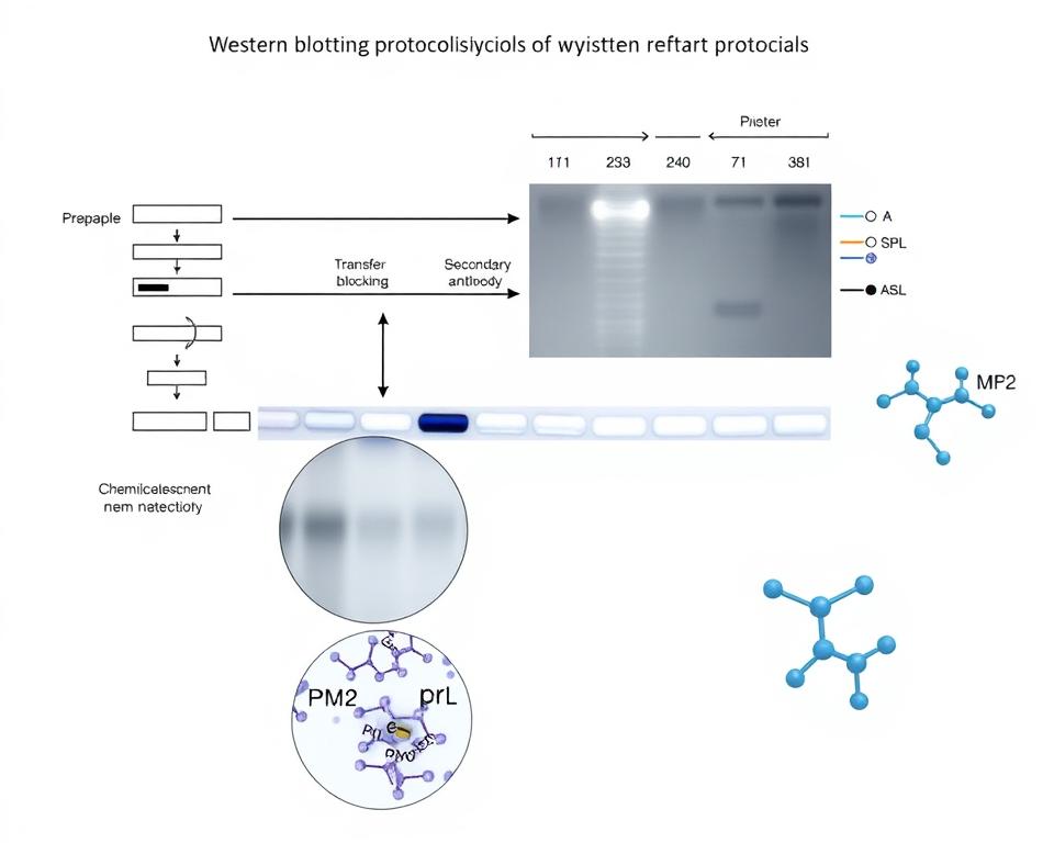
Leave a Comment
Your email address will not be published. Required fields are marked *