
Protein targeting research demands highly specific detection tools that deliver consistent results across multiple experimental platforms. We present a comprehensive characterization of a validated immunoreagent designed for investigating membrane-cytoskeleton dynamics in cellular systems.
Ezrin, also known as VIL2 or cytovillin, serves as a critical linker between cellular membranes and the cytoskeleton. This multidomain protein regulates essential processes including cell differentiation, adhesion, and migration. Understanding its function requires reliable detection methods for protein targeting applications.
The rabbit monoclonal approach offers significant advantages over traditional polyclonal alternatives. You gain superior specificity, consistent batch-to-batch performance, and renewable production capacity. These features make this technology ideal for long-term research projects requiring standardized results.
CPTC-Ezrin-2 has undergone extensive antibody characterization through multiple validation platforms. We confirmed its performance in immuno-MRM, Western blot, and ELISA applications. This validated reagent enables precise investigation of EZR protein expression patterns across diverse research contexts.
Key Takeaways
- Ezrin functions as a membrane-cytoskeleton linker protein essential for cell migration, adhesion, and differentiation processes
- Rabbit monoclonal antibodies provide superior specificity and batch-to-batch consistency compared to polyclonal alternatives
- CPTC-Ezrin-2 has been validated across multiple platforms including immuno-MRM, Western blot, and ELISA applications
- This immunoreagent enables reliable protein targeting investigations in cell biology and cancer research studies
- Comprehensive characterization data supports informed selection of detection tools for ezrin-related experiments
- The reagent addresses critical needs in metastatic cancer research where ezrin overexpression has significant implications
Introduction to Ezrin Rabbit Monoclonal Antibody
Understanding the molecular architecture of ezrin protein provides essential context for appreciating the value of monoclonal antibody technology in protein targeting research. The villin-2 antibody serves as a specialized tool for detecting and analyzing this critical structural protein across diverse experimental applications. We explore both the biological significance of the target protein and the technological advantages that make rabbit monoclonal antibodies superior research reagents.
Ezrin represents a fundamental component of cellular architecture that researchers worldwide investigate to understand disease mechanisms and normal physiological processes. The specificity and reliability of detection methods directly influence the quality of experimental outcomes in protein targeting studies.
Structural Organization and Biological Roles of Ezrin
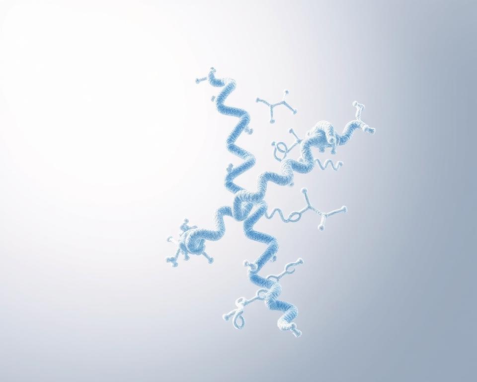
Ezrin belongs to the ERM protein family and functions as a membrane-cytoskeleton linker antibody target through its unique three-domain architecture. This modular organization enables ezrin to coordinate complex interactions between plasma membrane components and the underlying cytoskeletal framework.
The protein consists of three distinct structural regions that work together to regulate cellular processes:
- N-terminal FERM domain (N-ERMAD) – binds membrane proteins and phosphatidylinositol 4,5-bisphosphate
- Central α-helical domain – facilitates protein-protein interactions and regulatory functions
- C-terminal ERMAD – directly associates with filamentous actin structures
In resting cells, ezrin resides in an autoinhibited conformation where the N-terminal and C-terminal domains interact with each other. This dormant state prevents inappropriate activation and maintains cellular homeostasis.
Activation requires two coordinated molecular events. First, PtdIns(4,5)P2 binding disrupts the autoinhibited structure. Second, phosphorylation at threonine 567 stabilizes the open, active conformation that enables functional interactions.
The ezrin function encompasses multiple cellular processes essential for tissue organization and cell behavior. These include formation and maintenance of actin-rich surface structures such as microvilli, which increase membrane surface area for absorption and secretion. Ezrin also regulates cell-cell and cell-matrix contacts that determine tissue architecture.
Cell migration represents another critical aspect of ezrin function, particularly in developmental processes and cancer metastasis. Research demonstrates that ezrin expression increases significantly in highly metastatic cancers, making it a valuable biomarker and potential therapeutic target.
The membrane-cytoskeleton linker antibody targeting ezrin enables researchers to visualize protein localization during dynamic cellular processes. Signal transduction pathways frequently utilize ezrin as a scaffolding protein that brings signaling molecules into proximity with their substrates.
Advantages of Monoclonal Antibody Technology
Monoclonal antibodies represent a transformative advancement in protein detection methodology compared to traditional polyclonal sera. We recognize that research quality depends fundamentally on reagent consistency and specificity.
Rabbit monoclonal antibodies offer exceptional specificity through recognition of single epitopes on target proteins. This singular focus eliminates the signal variability inherent to polyclonal antibodies, which contain mixed populations recognizing multiple epitopes with varying affinities.
The renewable nature of monoclonal production ensures long-term availability of identical reagents. Immortalized cell lines generate consistent antibody batches indefinitely, eliminating batch-to-batch variation that compromises experimental reproducibility.
Rabbit hosts produce antibodies with distinct advantages over traditional mouse monoclonal systems. The rabbit immune system generates higher affinity antibodies with broader epitope recognition capabilities. This enhanced performance proves particularly valuable when detecting proteins in their native conformations.
Post-translational modifications require detection methods that distinguish subtle molecular differences. Rabbit monoclonal antibodies excel at recognizing phosphorylation states, glycosylation patterns, and other modifications critical to understanding protein regulation.
The combination of ERM protein family targeting specificity and rabbit monoclonal technology creates powerful research tools. These antibodies maintain consistent performance across multiple applications including immunohistochemistry, western blotting, and flow cytometry.
Research laboratories benefit from the documented validation that accompanies quality monoclonal antibodies. We emphasize that proper characterization data enables confident experimental design and reliable interpretation of results in protein targeting studies.
Key Characteristics of Ezrin Rabbit Monoclonal Antibody
Technical characterization data demonstrates how the p81 antibody achieves selective detection of ezrin protein through specific molecular recognition mechanisms. The CPTC-Ezrin-2 reagent exhibits well-defined binding properties that make it suitable for multiple experimental applications. We present comprehensive performance metrics that validate this antibody as a reliable tool for protein targeting studies.
The rabbit mAb ezrin targets a specific eight-amino-acid sequence within the ezrin protein structure. This antibody recognizes multiple forms of the target antigen across different experimental contexts. Performance validation includes quantitative binding measurements and detection capabilities in complex biological samples.
Molecular Recognition and Binding Performance
The foundation of antibody specificity lies in precise epitope recognition. CPTC-Ezrin-2 binds to Ezrin Peptide 1, which contains the sequence SGYLSSER. This eight-residue motif represents a unique epitope within the ezrin protein that enables selective detection without significant cross-reactivity to related ERM family members under optimized experimental conditions.
The target antigen carries multiple nomenclature designations in scientific literature. Researchers may encounter this protein under several aliases:
- EZR (primary gene symbol)
- VIL2 and Villin 2 (historical nomenclature)
- CVL, CVIL, and Cytovillin (functional descriptors)
- P81 (molecular weight designation)
- Villin-2 and Cytovillin 2 (alternative protein names)
Antibody affinity measurements provide quantitative assessment of binding strength. We evaluated CPTC-Ezrin-2 performance using indirect ELISA assays. The B50% metric represents the antibody concentration required to achieve 50% of maximum binding signal. High binding values in this assay indicate strong antibody-antigen interactions that support reliable detection across applications.
The versatility of epitope recognition extends across multiple protein presentations. CPTC-Ezrin-2 successfully detects synthetic peptides corresponding to the target sequence. Recombinant full-length ezrin protein expressed in bacterial expression systems shows positive recognition. Endogenous ezrin protein in mammalian cell lysates and tissue samples demonstrates consistent detection.
This multi-format recognition capability confirms epitope accessibility regardless of protein context. The antibody maintains binding performance whether the target appears as a short peptide fragment or within the complete three-dimensional protein structure. Such versatility proves essential for diverse experimental designs.
| Detection Method | Target Format | Result | Application Relevance |
|---|---|---|---|
| Indirect ELISA | Synthetic peptide | High B50% binding | Epitope mapping studies |
| Western blot | Recombinant protein | Positive detection | Expression validation |
| Immuno-MRM | Endogenous protein | Positive quantification | Proteomics analysis |
| Immunoprecipitation | Native complexes | Successful capture | Interaction studies |
Quantitative binding data supports the classification of this reagent as a high-affinity antibody. Equilibrium dissociation constants for similar ezrin-targeting molecules range from micromolar to nanomolar values. Lower dissociation constants indicate stronger binding, which translates to enhanced sensitivity in detection applications.
Immunoglobulin Class and Available Formats
The molecular classification of CPTC-Ezrin-2 identifies it as a rabbit monoclonal IgG. This immunoglobulin class represents the most abundant antibody type in circulation and offers optimal stability for laboratory applications. IgG molecules maintain functional integrity across various buffer conditions and storage temperatures.
The monoclonal production method ensures absolute consistency in epitope recognition. Every antibody molecule in the preparation targets the identical sequence with identical binding characteristics. This uniformity eliminates batch-to-batch variation that affects polyclonal antibody preparations.
We provide this antibody in purified immunoglobulin format suitable for direct experimental use. The purification process removes non-specific proteins while preserving antibody activity. Researchers can apply the reagent directly to immunohistochemistry, Western blotting, and flow cytometry protocols without additional preparation steps.
Concentration specifications enable precise experimental control. Standard working dilutions range from 1:500 to 1:2000 for most applications, though optimal concentrations require empirical determination for specific experimental systems. The high antibody affinity supports effective detection even at conservative dilutions, reducing reagent consumption while maintaining signal strength.
The rabbit monoclonal platform combines the specificity advantages of monoclonal antibodies with the high affinity characteristics of rabbit immune responses. Rabbits generate antibodies with longer complementarity-determining regions compared to mouse monoclonals. These extended binding surfaces create more extensive contacts with target epitopes, resulting in superior binding strength.
Reproducibility represents a critical advantage of monoclonal antibodies in protein targeting studies. Experiments conducted months or years apart yield comparable results when using the same monoclonal clone. This consistency proves essential for longitudinal studies and multi-center research collaborations where experimental standardization determines data validity.
Applications in Protein Targeting Studies
We have observed extensive utilization of the Ezrin Rabbit Monoclonal Antibody across various research contexts, from cellular imaging to pathway analysis. The antibody demonstrates remarkable versatility in contemporary protein targeting studies. Researchers employ this reagent to investigate ezrin behavior in multiple experimental designs.
The practical applications span several critical areas of cell biology research. Immunofluorescence applications enable detailed visualization of protein localization patterns within cells and tissues. These methodologies provide essential insights into ezrin distribution and function across diverse biological systems.
Modern protein targeting studies require reliable detection methods that maintain specificity across varied experimental conditions. The Ezrin Rabbit Monoclonal Antibody fulfills these requirements in numerous laboratory settings. Researchers benefit from consistent performance across multiple application platforms.
Cellular Localization Studies
Immunofluorescence protocols represent the primary method for mapping ezrin distribution within cellular compartments. We recommend using the antibody as a primary detection reagent followed by fluorophore-conjugated secondary antibodies. This combination enables high-resolution visualization through confocal or widefield fluorescence microscopy.
Research demonstrates that ezrin concentrates at the apical surface of polarized epithelial cells. The protein enriches particularly in microvilli and membrane ruffles where it cross-links actin filaments to membrane proteins. These protein localization patterns reveal fundamental aspects of cellular architecture and polarity maintenance.
Co-localization experiments provide additional insights into protein-protein interactions. Studies combining ezrin detection with S100P markers show these proteins associate in stimulated cells. Confocal laser scanning microscopy confirms this spatial relationship with exceptional clarity.
Dynamic cellular processes trigger ezrin redistribution that researchers can track using this antibody. During cell migration, ezrin accumulates at leading edge lamellipodia and trailing edge uropods. These observations illuminate the protein’s role in cytoskeletal reorganization during movement.
Multi-marker immunofluorescence studies combine ezrin visualization with other cellular components. Researchers frequently pair ezrin detection with actin staining or membrane receptor markers like CD44. These comprehensive immunofluorescence applications reveal functional protein complexes and their spatial organization.
Signal Transduction Pathway Analysis
Co-immunoprecipitation experiments leverage the antibody to capture ezrin from cellular lysates. Researchers then employ mass spectrometry or Western blot analysis to identify binding partners. This approach uncovers proteins involved in Rho GTPase signaling and phosphatidylinositol 3-kinase pathways.
The western blot ezrin antibody successfully detects the protein in cellular fractions with high sensitivity. You can employ this method to track ezrin expression across different experimental conditions. Western blotting provides quantitative data about protein abundance and post-translational modifications.
Monitoring ezrin phosphorylation reveals activation dynamics in response to extracellular stimuli. Growth factors, cytokines, and other signaling molecules trigger phosphorylation at specific residues. The western blot ezrin antibody enables researchers to distinguish between active and inactive protein pools.
Signal transduction studies benefit from the antibody’s compatibility with cellular fractionation techniques. We observe consistent detection across cytoplasmic, membrane, and cytoskeletal fractions. This capability allows comprehensive mapping of ezrin distribution during signaling events.
| Application Method | Primary Use | Detection Capability | Quantification |
|---|---|---|---|
| Immunofluorescence | Cellular visualization | Subcellular localization patterns | Semi-quantitative intensity analysis |
| Western Blotting | Protein expression analysis | Total and phosphorylated ezrin | Quantitative band densitometry |
| Co-immunoprecipitation | Protein interaction studies | Binding partner identification | Relative binding affinity assessment |
| Flow Cytometry | Population analysis | Cell surface expression levels | Quantitative fluorescence intensity |
Immune Response Investigations
Immunology research applications focus on ezrin function in leukocyte migration and lymphocyte activation. The antibody enables comprehensive analysis of immune cell behavior during physiological responses. Flow cytometry protocols provide quantitative measurements of ezrin expression across different immune cell populations.
Cell surface antigen detection capabilities make this antibody particularly valuable for immunology studies. Ezrin localizes to membrane domains where it regulates cell adhesion and motility. You can employ flow cytometry to measure surface-accessible ezrin on living cells without permeabilization.
Immune cell differentiation studies track ezrin expression changes during maturation processes. Western blotting reveals expression level variations as cells transition through developmental stages. These temporal expression patterns correlate with functional changes in cell migration capacity.
Lymphocyte activation triggers ezrin redistribution that researchers monitor using immunofluorescence techniques. The protein relocates to the immunological synapse during T cell receptor engagement. This dynamic behavior highlights ezrin’s participation in immune response coordination.
We recommend the antibody for investigating ezrin involvement in immune cell extravasation. During inflammation, leukocytes must cross endothelial barriers to reach tissue sites. Ezrin facilitates this process through membrane-cytoskeleton reorganization that the antibody helps visualize.
Cell surface antigen detection applications extend to studying ezrin expression on different immune cell subsets. Natural killer cells, macrophages, and dendritic cells exhibit varied ezrin levels that correlate with functional specialization. Flow cytometry sorting combined with Western blot validation provides comprehensive characterization across immune compartments.
The antibody’s performance in cell surface antigen detection protocols enables real-time monitoring of ezrin availability at plasma membranes. This capability proves essential for understanding how cells regulate membrane protein organization during immune responses. Researchers gain insights into the molecular mechanisms underlying immune cell function and dysfunction.
Comparing Ezrin Rabbit Monoclonal Antibody with Alternatives
Antibody comparison begins with evaluating how different detection reagents perform in ezrin protein targeting applications. We recognize that researchers must consider multiple factors when selecting the optimal tool for their experimental needs. The cytovillin antibody landscape includes both traditional polyclonal preparations and various monoclonal formats, each offering distinct characteristics.
Understanding antibody selection criteria helps you make informed decisions that directly impact experimental reproducibility and data quality. Different reagent types present unique advantages and limitations based on your specific research applications. The following analysis examines key alternatives to guide your selection process.
Polyclonal Antibodies for Ezrin Detection
Traditional polyclonal antibodies represent the historical approach to ezrin protein detection. These reagents contain heterogeneous antibody populations that recognize multiple epitopes across the target protein. The multi-epitope recognition pattern can provide robust signal amplification in certain experimental conditions.
Polyclonal preparations demonstrate tolerance for epitope masking and protein conformational variations. This flexibility proves valuable when working with native protein structures or partially denatured samples. The diverse antibody population increases the likelihood that some molecules will bind successfully even under challenging conditions.
However, the polyclonal versus monoclonal debate centers on critical limitations of polyclonal reagents. Batch-to-batch variability represents the primary disadvantage affecting experimental reproducibility. Each new production lot contains different antibody populations with varying specificities and affinities.
We observe that polyclonal antisera may include antibodies recognizing unintended targets, increasing background signal and reducing specificity. Once the immunized animal source is depleted, you cannot reproduce the exact reagent composition. This limitation requires revalidation when switching lots and creates long-term supply chain concerns.
Monoclonal formats eliminate these reproducibility challenges through consistent performance across unlimited production runs. Every antibody molecule from a monoclonal clone recognizes the identical epitope with uniform affinity. This ensures that results obtained today remain reproducible years later using the same clone.
Alternative Rabbit Monoclonal Options
Various rabbit monoclonal clones targeting ezrin recognize distinct epitopes within the protein sequence. Some clones target the N-terminal FERM domain, while others bind the central region or C-terminal domain. Epitope location significantly influences application suitability and detection reliability.
N-terminal epitopes remain accessible in both full-length ezrin and truncated protein forms. C-terminal epitopes may become masked in certain protein conformations or when ezrin engages in binding interactions. Understanding these epitope-dependent characteristics helps you select the cytovillin antibody variant best suited for your experimental design.
Rabbit monoclonal antibodies generally demonstrate higher affinity compared to mouse monoclonals. This advantage stems from differences in immune system repertoire and affinity maturation processes between species. The enhanced binding strength translates to improved sensitivity in detection applications.
| Characteristic | Polyclonal Antibodies | Rabbit Monoclonals | Mouse Monoclonals |
|---|---|---|---|
| Epitope Recognition | Multiple epitopes | Single epitope | Single epitope |
| Batch Consistency | Variable between lots | Identical across production | Identical across production |
| Binding Affinity | Mixed population | High (nM to pM range) | Moderate (nM range) |
| Background Signal | Higher potential | Lower due to specificity | Lower due to specificity |
| Long-term Availability | Limited by animal source | Unlimited from cell line | Unlimited from cell line |
When evaluating different rabbit monoclonal alternatives, you should prioritize validated applications and characterized specificity data. Documented performance in techniques like immunohistochemistry, Western blotting, or flow cytometry provides confidence in reagent suitability. Independent validation studies offer additional assurance beyond manufacturer claims.
Epitope mapping information helps predict antibody performance in your specific experimental context. Antibodies targeting structured domains may require native protein conditions, while those recognizing linear epitopes work well with denatured samples. Applying proper antibody selection criteria based on these technical considerations optimizes your experimental outcomes and ensures reproducible protein targeting results.
Protocols for Utilizing Ezrin Rabbit Monoclonal Antibody
We provide comprehensive protocol guidance to help you maximize the performance of ezrin rabbit monoclonal antibody in your research applications. Proper implementation across different detection methods requires understanding the specific technical requirements and optimization strategies for each platform. This section delivers practical protocols that enable researchers to achieve consistent, reliable results in protein targeting studies.
The success of your experiments depends on selecting appropriate detection methods and following validated procedures. We outline three primary application scenarios where ezrin rabbit monoclonal antibody demonstrates proven utility: immunohistochemistry for tissue localization, western blotting for protein expression analysis, and flow cytometry for quantitative cell population studies. Each methodology presents unique technical considerations that influence antibody performance and data quality.
Tissue-Based Detection Approaches
Immunohistochemistry techniques enable visualization of ezrin protein distribution within fixed tissue sections and cultured cells. Standard immunohistochemistry ezrin protocols begin with proper tissue preparation, typically using formalin-fixed paraffin-embedded specimens. You should deparaffinize sections using xylene or xylene substitutes, followed by rehydration through graded alcohol series.
Antigen retrieval represents a critical step for exposing epitopes masked during fixation. Heat-induced epitope retrieval using citrate buffer (pH 6.0) or EDTA buffer (pH 8.0) in a pressure cooker or steamer for 20 minutes typically yields optimal results. Allow slides to cool gradually to room temperature before proceeding to blocking steps.
Blocking non-specific binding sites improves signal-to-noise ratio significantly. Apply blocking solution containing 5-10% normal serum from the secondary antibody host species combined with bovine serum albumin for 60 minutes at room temperature. This step prevents background staining that can complicate interpretation of ezrin localization patterns.
Primary antibody incubation requires careful attention to antibody dilution and incubation conditions. We recommend starting with dilutions ranging from 1:100 to 1:1000, with overnight incubation at 4°C typically providing superior results compared to shorter room temperature incubations. Optimize the dilution empirically using positive control tissues such as normal intestinal epithelium, where ezrin concentrates in enterocyte microvilli.
Detection methods for immunohistochemistry include both chromogenic and fluorescent approaches. Fluorophore-conjugated secondary antibodies enable multiplexing capabilities, while enzyme-conjugated antibodies with DAB substrate offer permanent staining suitable for long-term archival. Always include negative controls by omitting primary antibody to validate staining specificity.
Researchers should note that characterization data indicates variable performance in immunohistochemistry applications for certain ezrin antibody clones. The CPTC-Ezrin-2 clone showed negative results in standard IHC evaluation, highlighting the importance of validation in your specific tissue type and fixation conditions before undertaking large-scale studies.
Lysate-Based Protein Detection
Western blotting applications provide quantitative assessment of ezrin expression levels in cell and tissue lysates. The western blot ezrin antibody demonstrates validated performance for detecting the approximately 80 kDa ezrin protein, sometimes designated as p81. Begin by preparing high-quality protein lysates using appropriate lysis buffers.
Sample preparation requires lysis buffer containing detergents such as Triton X-100 or NP-40 at concentrations of 0.5-1%. Include protease inhibitor cocktails to prevent protein degradation during extraction. If you plan to analyze ezrin phosphorylation states, add phosphatase inhibitors such as sodium orthovanadate and sodium fluoride to preserve post-translational modifications.
Quantify protein concentrations using Bradford or BCA assays to ensure equal loading across lanes. Load 20-50 micrograms of total protein per lane for most applications, though optimization may be necessary based on ezrin expression levels in your specific sample type. Include molecular weight markers spanning 50-100 kDa for accurate size determination.
SDS-PAGE separation using 8-12% polyacrylamide gels resolves ezrin effectively from other proteins. Transfer separated proteins to PVDF or nitrocellulose membranes using wet, semi-dry, or dry transfer systems according to manufacturer specifications. PVDF membranes generally provide higher binding capacity and durability for reprobing experiments.
Block membranes with 5% non-fat dry milk or 3-5% BSA in Tris-buffered saline with 0.1% Tween-20 (TBST) for one hour. Incubate with western blot ezrin antibody at validated antibody dilution ratios, typically 1:500 to 1:2000, overnight at 4°C with gentle agitation. Wash membranes thoroughly with TBST before applying secondary antibodies.
Secondary antibodies conjugated to horseradish peroxidase enable chemiluminescent detection, while fluorophore-conjugated secondaries support multiplexing with other target proteins. Use enhanced chemiluminescence reagents and appropriate imaging systems to capture band intensity data. Include positive control lysates from cells known to express ezrin abundantly, such as A431 or HeLa cell lines.
| Application Method | Recommended Dilution | Incubation Conditions | Expected Results |
|---|---|---|---|
| Immunohistochemistry | 1:100 to 1:1000 | Overnight at 4°C or 2 hours at RT | Membrane/cytoplasmic localization |
| Western Blotting | 1:500 to 1:2000 | Overnight at 4°C | Single band at 80 kDa |
| Flow Cytometry | 1:50 to 1:200 | 30-60 minutes at 4°C | Quantitative expression profiles |
| Immunofluorescence | 1:100 to 1:500 | 1-2 hours at RT | Cytoskeletal and membrane staining |
Quantitative Cell Analysis Methods
Flow cytometry protocols enable quantitative measurement of ezrin expression across cell populations. This approach proves particularly valuable for analyzing immune cells or cancer cell lines where ezrin levels correlate with metastatic potential and invasive behavior. Flow cytometry provides single-cell resolution that reveals expression heterogeneity within populations.
Prepare single-cell suspensions by enzymatic digestion or mechanical dissociation, maintaining cell viability above 90% for optimal results. Fix cells using 4% formaldehyde in phosphate-buffered saline for 10-15 minutes at room temperature. Permeabilize cells with 0.1-0.5% Triton X-100 for 5-10 minutes to allow antibody access to intracellular ezrin pools.
Block non-specific binding with 2-5% BSA or normal serum for 30 minutes before antibody staining. Apply ezrin rabbit monoclonal antibody at dilutions between 1:50 to 1:200 for 30-60 minutes at 4°C. The detection methods for flow cytometry require fluorophore-conjugated secondary antibodies compatible with your instrument’s laser and filter configurations.
Analyze stained samples promptly or fix in 1% paraformaldehyde if delayed acquisition is necessary. Gate on viable, single cells using forward and side scatter parameters. Include unstained controls, isotype controls, and fluorescence-minus-one controls to establish gating strategies and distinguish positive populations from background fluorescence. These flow cytometry protocols deliver reproducible quantitative data suitable for comparative expression studies.
Case Studies Utilizing Ezrin Rabbit Monoclonal Antibody
Documented research investigations reveal how ezrin-targeted antibodies contribute to breakthrough discoveries in cancer progression and tissue formation. We examine compelling examples that demonstrate the practical value of the Ezrin Rabbit Monoclonal antibody across diverse scientific disciplines. These real-world applications showcase how precise protein detection advances our understanding of disease mechanisms and normal biological processes.
The antibody has enabled researchers to uncover critical molecular pathways that were previously difficult to visualize. Scientists now track ezrin expression patterns with confidence, generating reproducible data that shapes therapeutic development strategies.
Oncological Research Applications
Investigations into cancer metastasis research have revealed ezrin as a pivotal player in tumor progression. Studies focusing on non-small cell lung carcinomas demonstrate that ezrin overexpression correlates directly with metastatic potential. Researchers use the Ezrin Rabbit Monoclonal antibody to assess expression levels across different tumor grades, establishing ezrin as both a biomarker and therapeutic target.
Malignant gliomas represent another area where ezrin detection has provided critical insights. Research demonstrates that ezrin is required for tumor cell invasion through three-dimensional matrices. Scientists employ immunofluorescence techniques to visualize ezrin redistribution to invasive protrusions at the leading edge of migrating cancer cells.
The antibody facilitates investigation of activation states through detection of phosphorylated forms. Phospho-threonine 567 represents the active conformation, and tracking this modification reveals when ezrin transitions to its functional state. Western blot analyses track expression changes during epithelial-mesenchymal transition, the process by which epithelial tumor cells acquire migratory properties.
Co-immunoprecipitation studies using ezrin antibodies have identified critical binding partners. S100P-mediated activation of ezrin promotes transendothelial migration, a key step in metastatic dissemination. This discovery emerged from protein expression studies that mapped ezrin interaction networks within tumor microenvironments.
Receptor tyrosine kinase research has uncovered additional complexity in ezrin regulation. Tyrosine phosphorylation of ezrin following Kit and Flt3 receptor activation demonstrates metastasis-promoting functions. These findings emerged through experiments combining receptor stimulation with ezrin detection protocols.
Functional assays paired with ezrin detection reveal mechanistic details. Studies show that ezrin depletion or mutation impairs tumor cell invasion and reduces metastatic colonization in experimental models. The antibody enables researchers to correlate protein levels with functional outcomes across multiple cancer types.
Embryonic and Tissue Development Research
Research utilizing the antibody to investigate ezrin in development has illuminated fundamental processes in tissue morphogenesis. Scientists track protein expression during embryonic development, particularly in tissues undergoing epithelial polarization. The developing intestine, kidney tubules, and neural tube show distinct ezrin localization patterns that guide cellular organization.
Immunofluorescence studies reveal ezrin accumulation at the apical membrane domain where it organizes microvilli formation. This spatial distribution is essential for establishing apical-basal polarity in epithelial cells. The antibody allows researchers to visualize how ezrin contributes to morphogenetic movements during gastrulation and organogenesis through regulation of cell shape changes.
Developmental immunology investigations examine ezrin expression during lymphocyte maturation. ERM proteins are required for efficient leukocyte migration, and protein expression studies demonstrate dynamic regulation throughout immune cell development. Researchers use the antibody to track ezrin levels as immune cells acquire migratory capabilities necessary for tissue surveillance.
The antibody facilitates comparative analyses across developmental stages. Scientists document temporal expression patterns that correlate with specific morphological transitions. These longitudinal studies establish ezrin as a key regulator of cellular architecture during tissue formation.
| Research Application | Biological System | Key Findings | Detection Method |
|---|---|---|---|
| Metastatic progression | Non-small cell lung carcinoma | Overexpression correlates with invasion potential | Immunohistochemistry, Western blot |
| Glioma invasion | Malignant brain tumors | Required for Rho/ROCK-dependent motility | Immunofluorescence, functional assays |
| Epithelial polarity | Developing intestine and kidney | Establishes apical membrane organization | Immunofluorescence microscopy |
| Immune cell migration | Lymphocyte maturation | Essential for efficient tissue homing | Flow cytometry, Western blot |
These case studies demonstrate the versatility and reliability of the Ezrin Rabbit Monoclonal antibody across varied research contexts. From cancer metastasis research to developmental investigations, the antibody provides consistent, specific detection that enables meaningful scientific discoveries. Researchers continue to expand applications as new questions about ezrin function emerge in both pathological and physiological settings.
Challenges and Considerations
Understanding potential obstacles in antibody performance is essential for researchers working with EZR antibody in diverse experimental contexts. Technical challenges can arise from protein family similarities, application-specific requirements, and sample preparation variables. We provide systematic guidance to help you navigate these considerations and achieve consistent, reliable results in your protein targeting studies.
Addressing these challenges proactively through antibody validation and methodical optimization improves experimental reproducibility. Researchers benefit from understanding both the molecular basis of potential complications and practical troubleshooting protocols. This knowledge enables informed decision-making when designing experiments and interpreting results.
Specificity Verification in ERM Protein Family Context
The ERM protein family presents unique specificity considerations for EZR antibody applications. Ezrin, radixin, and moesin share substantial sequence homology, particularly within conserved FERM domains and C-terminal actin-binding regions. This structural similarity creates potential for cross-reactivity if the antibody epitope corresponds to a conserved sequence motif across family members.
While the Ezrin Rabbit Monoclonal Antibody targets a peptide sequence designed specifically for ezrin, validation in your experimental system remains critical. This becomes especially important when working with tissues or cell types expressing multiple ERM family members simultaneously. We recommend implementing comprehensive validation strategies before drawing experimental conclusions.
Several validation approaches provide confidence in antibody specificity. Western blot analysis using recombinant purified ezrin, radixin, and moesin proteins directly assesses cross-reactivity potential. This method allows side-by-side comparison of antibody binding to each family member under identical conditions.
Genetic manipulation strategies offer functional validation of antibody specificity. siRNA or CRISPR-mediated knockdown of ezrin followed by immunodetection should result in signal disappearance if the antibody is truly specific. The presence of remaining signal after ezrin depletion suggests cross-reactivity with other proteins.
Peptide competition assays provide additional validation through epitope-specific blocking. Pre-incubation of the EZR antibody with its immunizing peptide should prevent specific binding to ezrin. Signals that persist after peptide blocking may indicate non-specific interactions requiring further optimization.
Post-translational modifications near the epitope region can influence antibody recognition. Phosphorylation, ubiquitination, or other modifications may alter epitope accessibility or antibody binding affinity. Researchers should consider these variables when interpreting results, particularly in signaling pathway studies where ezrin phosphorylation status changes dynamically.
Systematic Approaches to Method Refinement
Application-specific optimization is necessary because antibodies often perform differently across techniques. Detection optimization requirements vary between Western blot, immunofluorescence, and immunohistochemistry due to differences in protein denaturation states and epitope accessibility. You may need to adjust protocols for each application independently.
Weak or absent signals require systematic troubleshooting to identify the limiting factor. Serial dilution testing helps determine optimal antibody concentration for your specific samples. Extended incubation times may improve binding kinetics, particularly when working with low-abundance targets or challenging tissue samples.
Enhanced blocking strategies reduce background while preserving specific signal. Testing different blocking agents—including BSA, non-fat milk, or synthetic blockers—identifies the most effective option for your application. Signal amplification systems provide alternatives when target abundance limits detection with standard protocols.
High background signals necessitate optimization of washing and blocking conditions. Increased washing stringency with higher salt concentrations or additional wash steps removes non-specifically bound antibody. Reducing antibody concentration often improves signal-to-noise ratio when background exceeds acceptable levels.
Sample preparation critically influences antibody validation outcomes and experimental reproducibility. Maintaining appropriate protease inhibitors in lysis buffers prevents ezrin degradation during sample processing. Avoiding excessive freeze-thaw cycles preserves protein integrity and epitope structure for optimal antibody recognition.
Adequate sample loading ensures sufficient target protein for detection across different applications. We recommend optimizing protein input amounts through preliminary titration experiments. This approach establishes baseline parameters for consistent results across experimental replicates.
Fixation method selection significantly impacts immunofluorescence and immunohistochemistry results. Paraformaldehyde fixation preserves cellular morphology while maintaining reasonable epitope accessibility. Methanol fixation provides enhanced membrane permeability but may alter protein conformation and reduce antibody binding.
The following table compares validation methods and optimization strategies for troubleshooting protocols:
| Validation Method | Application | Key Advantage | Technical Consideration |
|---|---|---|---|
| Recombinant Protein Testing | Western Blot | Direct assessment of cross-reactivity with ERM family members | Requires access to purified ezrin, radixin, and moesin proteins |
| Genetic Knockdown Validation | All Applications | Functional confirmation of antibody specificity in cellular context | Time-intensive process requiring molecular biology expertise |
| Peptide Competition Assay | Immunofluorescence, IHC | Epitope-specific blocking confirms binding specificity | Effective only when immunizing peptide is accessible |
| Serial Dilution Optimization | All Applications | Identifies optimal antibody concentration for signal-to-noise ratio | Requires multiple experimental iterations for each application |
Temperature and incubation conditions influence antibody binding kinetics and specificity. Room temperature incubations may reduce background compared to higher temperatures, though they may also decrease binding efficiency. Overnight incubations at 4°C often provide optimal results for detection optimization in immunofluorescence applications.
Buffer composition affects antibody stability and binding characteristics. Tris-buffered saline versus phosphate-buffered saline can influence background levels and specific signal intensity. We recommend testing both buffer systems when establishing new protocols or troubleshooting unexpected results.
Documentation of optimization parameters enables reproducibility across experiments and research teams. Recording antibody dilutions, incubation times, blocking conditions, and washing protocols creates reference standards for future work. This systematic approach to troubleshooting protocols accelerates experimental progress and reduces variability.
Future Directions in Ezrin Antibody Research
Recent advances in antibody engineering open new pathways for ezrin-based diagnostics and treatment strategies. The accumulated knowledge about ezrin’s role in cellular processes now drives innovation in both therapeutic development and research tool optimization. We see emerging opportunities that bridge fundamental research with clinical applications.
The transition from research reagent to therapeutic agent represents a significant evolution in ezrin antibody development. Evidence gathered through protein targeting studies positions ezrin as a valuable target for intervention in multiple disease contexts. These advances promise to expand the impact of rabbit mAb ezrin beyond traditional laboratory applications.

Therapeutic Opportunities for Ezrin Targeting
The documented role of ezrin in cancer metastasis creates compelling opportunities for therapeutic intervention. Studies using validated antibodies have identified ezrin overexpression in aggressive tumor types, establishing it as a potential treatment target. Several strategic approaches are under active investigation to exploit this vulnerability.
Antibody-drug conjugates represent a promising direction for ezrin-targeted therapy. These engineered molecules combine the specificity of ezrin antibodies with potent cytotoxic payloads. The conjugates selectively deliver chemotherapy agents to ezrin-overexpressing cancer cells, reducing systemic toxicity while maximizing tumor impact.
Bispecific antibody designs offer another innovative approach. These therapeutic antibodies simultaneously engage ezrin on tumor cells and immune effector molecules on T cells or NK cells. This dual targeting redirects the patient’s immune system to recognize and eliminate ezrin-positive cancer cells.
Small molecule inhibitors that disrupt ezrin interactions with binding partners like PtdIns(4,5)P2 are advancing through preclinical development. These compounds interfere with ezrin’s membrane-cytoskeleton linking function, potentially reducing metastatic capacity. Peptide-based interventions targeting specific protein interaction domains provide additional therapeutic modalities.
The application of ezrin antibodies in precision medicine extends beyond direct therapeutic use. Companion diagnostic development utilizes standardized immunohistochemistry assays to identify patients with high ezrin expression. This stratification approach guides treatment decisions, ensuring that ezrin-targeted therapies reach the patients most likely to benefit.
Beyond oncology, ezrin targeting shows potential in inflammatory diseases. The protein’s role in leukocyte migration suggests that modulating ezrin function could reduce pathological immune cell trafficking. Research continues to explore these applications across diverse disease contexts.
Advanced Production and Engineering Methods
Recombinant antibody production technologies have transformed how we generate research and therapeutic antibodies. Next-generation platforms utilize improved immunization strategies and high-throughput screening systems to identify optimal clones. These advances ensure consistent quality and eliminate batch-to-batch variability.
Animal-free production systems represent a significant technical advancement. Defined recombinant expression platforms produce antibodies with reproducible characteristics, addressing key concerns in both research reproducibility and therapeutic manufacturing. These systems also facilitate scalability for commercial applications.
Antibody engineering techniques expand the functional capabilities of ezrin-targeting reagents. Humanization reduces immunogenicity for therapeutic applications, while affinity maturation through directed evolution enhances binding strength. Format engineering creates specialized variants including single-chain variable fragments and diabodies with improved tissue penetration.
Computational antibody design using artificial intelligence accelerates development timelines. Machine learning algorithms predict optimal antibody sequences based on structural data and binding requirements. These predictive tools reduce the experimental burden of traditional antibody discovery.
DNA-encoded antibody libraries vastly expand candidate diversity for screening. This technology enables rapid identification of high-affinity binders from libraries containing billions of variants. The approach complements traditional hybridoma and phage display methods.
| Therapeutic Strategy | Mechanism of Action | Primary Application | Development Stage |
|---|---|---|---|
| Antibody-Drug Conjugates | Targeted cytotoxic delivery to ezrin-positive cells | Metastatic cancer treatment | Preclinical optimization |
| Bispecific Antibodies | Immune cell redirection against ezrin-expressing tumors | Immuno-oncology therapy | Early development |
| Companion Diagnostics | Patient stratification via ezrin expression profiling | Precision medicine guidance | Validation studies |
| Small Molecule Inhibitors | Disruption of ezrin-membrane interactions | Anti-metastatic therapy | Lead optimization |
The integration of advanced antibody engineering with recombinant antibody production creates reagents with unprecedented specificity and consistency. These technical improvements benefit both fundamental research applications and therapeutic development. The enhanced quality of modern rabbit mAb ezrin tools enables more reliable experimental results and supports regulatory requirements for clinical use.
Emerging production methods also address sustainability concerns. Synthetic biology approaches reduce reliance on animal immunization while maintaining antibody quality. These innovations align with evolving ethical standards and regulatory expectations in both research and pharmaceutical industries.
The convergence of computational design, high-throughput screening, and precision engineering positions ezrin antibody research at the forefront of molecular targeting innovation. We anticipate continued rapid progress as these technologies mature and integrate. The next generation of ezrin-targeting tools will offer researchers and clinicians more powerful options for investigating and intervening in ezrin-mediated processes.
Conclusion: The Impact of Ezrin Rabbit Monoclonal Antibody
The Ezrin Rabbit Monoclonal antibody stands as a validated membrane-cytoskeleton linker antibody that advances protein targeting research across multiple scientific disciplines. Its characterized performance in ELISA, Western blot, and immuno-MRM platforms provides researchers with confidence in experimental outcomes.
Validated Performance Across Research Applications
We have established that this antibody delivers specific recognition of ezrin while distinguishing it from related ERM family members. The tool enables investigation spanning basic cell biology studies to translational cancer research. Researchers examining cellular localization, signal transduction pathways, and immune responses benefit from the reproducibility inherent in monoclonal antibody applications.
Successful implementation requires attention to protocol optimization and appropriate validation controls. The guidance provided throughout this characterization supports diverse experimental designs and detection methods.
Expanding Research Horizons
Growing understanding of ezrin biology opens new directions for scientific advancement. Integration with advanced imaging techniques will reveal nanoscale organization patterns. Systems biology approaches combining antibody-based detection with proteomics will map comprehensive regulatory networks.
Standardized ezrin assays may transition into clinical laboratory settings as companion diagnostics. This exemplifies how research tools evolve into patient care applications. The continued development of refined detection reagents will support the expanding community investigating membrane-cytoskeleton interactions, providing precision instruments necessary for mechanistic discovery and therapeutic development in protein targeting research.
References and further readings:
1.Gautreau A, Louvard D, Arpin M. Morphogenic effects of ezrin require a phosphorylation-induced transition from oligomers to monomers at the plasma membrane. Journal of Cell Biology. 2000;150(1):193–203.
https://rupress.org/jcb/article-abstract/150/1/193/47737/Morphogenic-Effects-of-Ezrin-Require-a?redirectedFrom=fulltext2.Heiska L, Alfthan K, Grönholm M, Vilja P, Vaheri A, Carpén O. Association of ezrin with intercellular adhesion molecule-1 and -2 (ICAM-1 and -2): regulation by phosphatidylinositol 4,5-bisphosphate. Journal of Biological Chemistry. 1998;273(34):21893–21900.
https://www.jbc.org/article/S0021-9258(18)48863-1/fulltext3.Bretscher A, Edwards K, Fehon RG. ERM proteins and merlin: integrators at the cell cortex. Nature Reviews Molecular Cell Biology. 2002;3(8):586–599.
https://www.nature.com/articles/nrm8824.Fievet BT, Gautreau A, Roy C, Del Maestro L, Mangeat P, Louvard D, Arpin M. Phosphoinositide binding and phosphorylation act sequentially in the activation mechanism of ezrin. Journal of Cell Biology. 2004;164(5):653–659.
https://rupress.org/jcb/article-abstract/164/5/653/33737/Phosphoinositide-binding-and-phosphorylation-act?redirectedFrom=fulltext
FAQ
What is ezrin protein and why is it important in biological research?
Ezrin, also known as villin-2 or cytovillin, is a member of the ERM (ezrin/radixin/moesin) protein family that functions as a membrane-cytoskeleton linker. This protein features a three-domain structure including an N-terminal FERM domain that binds membrane proteins and phospholipids, a central α-helical region for protein-protein interactions, and a C-terminal domain that binds F-actin. Ezrin plays critical roles in establishing cellular polarity, forming microvilli, regulating cell migration and adhesion, and facilitating signal transduction pathways. Its significance in research stems from its involvement in both normal physiological processes and pathological conditions, particularly cancer metastasis where ezrin overexpression correlates with invasive potential in various malignancies including non-small cell lung carcinomas and osteosarcomas.
What advantages does the Ezrin Rabbit Monoclonal Antibody offer compared to polyclonal antibodies?
The rabbit mAb ezrin provides several critical advantages over traditional polyclonal antisera. Monoclonal antibodies offer exceptional specificity through recognition of a single epitope, ensuring consistent results across experiments and eliminating the batch-to-batch variation inherent to polyclonal sera where different production lots contain varying antibody populations. The renewable nature of monoclonal production from immortalized cell lines ensures long-term availability of identical reagents, whereas polyclonal antibodies cannot be reproduced once the immunized animal is depleted. Additionally, rabbit monoclonal antibodies demonstrate higher affinity and broader epitope recognition compared to mouse monoclonals, making them particularly valuable for detecting proteins in their native conformations and various post-translational modification states. This reproducibility and consistency are essential for reliable protein targeting studies.
Which applications have been validated for the Ezrin Rabbit Monoclonal Antibody?
The EZR antibody has demonstrated validated performance across multiple protein targeting applications. In Western blot applications, the antibody successfully detects ezrin at approximately 80 kDa (hence the designation p81) in cell and tissue lysates, enabling quantitative analysis of expression levels. For immunofluorescence studies, the antibody facilitates visualization of ezrin distribution in cells and tissues, revealing enrichment at apical surfaces of polarized epithelial cells, particularly in microvilli and membrane ruffles. The antibody enables co-immunoprecipitation experiments to identify ezrin binding partners and investigate protein-protein interactions. In flow cytometry applications, it allows quantification of ezrin expression levels in different cell populations. We note that characterization data indicates variable performance in immunohistochemistry applications for certain ezrin antibody clones, emphasizing the importance of application-specific validation in your experimental system.
What epitope does the Ezrin Rabbit Monoclonal Antibody recognize?
The Ezrin Rabbit Monoclonal Antibody recognizes a specific epitope within the ezrin protein sequence (SGYLSSER from Ezrin Peptide 1), ensuring highly selective detection. This epitope-specific recognition provides discrimination from closely related ERM family members radixin and moesin under optimized conditions, despite the substantial sequence homology these proteins share. The antibody successfully recognizes multiple forms of the target including synthetic peptides, recombinant full-length ezrin protein expressed in bacterial systems, and endogenous ezrin in mammalian cell lysates and tissue samples. This versatility confirms that the epitope remains accessible across different protein contexts, supporting diverse experimental applications from basic biochemical characterization to complex cellular studies.
How can I validate that the antibody is detecting ezrin specifically and not cross-reacting with related proteins?
We recommend several validation approaches to confirm specificity of the cytovillin antibody. First, perform Western blot analysis using recombinant purified ezrin, radixin, and moesin proteins to directly assess cross-reactivity with other ERM family members. Second, conduct siRNA or CRISPR-mediated knockdown of ezrin in your cell system, followed by immunodetection to confirm that the detected band disappears when ezrin is depleted. Third, implement peptide competition assays where pre-incubation of the antibody with the immunizing peptide should block specific binding to the target. For immunofluorescence applications, include appropriate negative controls (omitting primary antibody) and positive control tissues such as normal intestinal epithelium where ezrin is highly expressed in enterocyte microvilli. These validation steps ensure that your detected signal represents authentic ezrin protein rather than cross-reactive targets.
What is the recommended dilution range for Western blot applications?
For western blot ezrin antibody applications, typical dilution ranges fall between 1:1000 to 1:5000, though optimal dilution depends on antibody concentration, target protein abundance in your samples, and your detection system sensitivity. We recommend performing a dilution series starting at 1:500, 1:1000, 1:2000, and 1:5000 to identify the dilution that provides strong specific signal with minimal background in your particular experimental system. Ensure sample preparation includes appropriate lysis buffer composition with detergents (Triton X-100 or NP-40), protease inhibitors, and phosphatase inhibitors to preserve ezrin and its phosphorylation states. Include positive control lysates from cells known to express ezrin abundantly (such as epithelial cell lines) and molecular weight markers to confirm the expected ~80 kDa band. Primary antibody incubation typically occurs overnight at 4°C with gentle agitation for optimal results.
Can the Ezrin Rabbit Monoclonal Antibody detect phosphorylated forms of ezrin?
The Ezrin Rabbit Monoclonal Antibody recognizes total ezrin protein and can detect both non-phosphorylated and phosphorylated forms, provided the epitope (SGYLSSER sequence) is not directly modified by phosphorylation. This allows investigation of ezrin expression levels across different cellular states. However, for specific detection of activated ezrin phosphorylated at threonine 567—the key activation site that represents the functionally active conformation—you would require a phospho-specific antibody designed to recognize only the phosphorylated epitope. When using the total ezrin antibody in conjunction with appropriate sample preparation including phosphatase inhibitors, you preserve all phosphorylation states in your samples. This enables downstream analysis through techniques such as co-immunoprecipitation followed by phospho-detection using phospho-specific antibodies, or comparative Western blot analysis of samples treated with phosphatase inhibitors versus phosphatases to assess the proportion of phosphorylated ezrin.
What role does ezrin play in cancer metastasis, and how can the antibody support related research?
Ezrin has emerged as a significant protein in cancer biology due to its overexpression in metastatic cancers and functional contribution to tumor cell invasion and migration capabilities. Research using ezrin antibodies has demonstrated that elevated ezrin expression correlates with metastatic potential across various malignancies. Ezrin actively promotes metastasis by facilitating tumor cell invasion through three-dimensional matrices, enhancing transendothelial migration (a key step in metastatic dissemination), and regulating cell surface receptor localization. The membrane-cytoskeleton linker antibody supports cancer research through multiple approaches: Western blot analysis tracks ezrin expression changes during epithelial-mesenchymal transition; immunofluorescence reveals ezrin redistribution to invasive protrusions at leading edges of migrating cancer cells; co-immunoprecipitation identifies critical binding partners such as S100P whose interaction with ezrin promotes metastatic behavior; and immunohistochemistry on tumor specimens correlates ezrin levels with clinical outcomes, supporting its potential as both a biomarker and therapeutic target.
How should I optimize the antibody if I’m experiencing high background or weak signal?
For optimizing detection methods with the EZR antibody, we recommend systematic troubleshooting approaches tailored to your specific challenge. For weak or absent signals: perform serial dilution testing from 1:100 to 1:5000 to identify optimal antibody concentration; extend primary antibody incubation time to overnight at 4°C; implement signal amplification systems; increase protein loading amounts; and verify that your samples contain adequate ezrin expression using positive control cell lines or tissues. For high background signals: increase washing stringency with additional washes or higher salt concentrations in wash buffers; optimize blocking conditions by testing different blocking agents (BSA, non-fat milk, or synthetic blockers); reduce primary antibody concentration; decrease incubation time; and ensure proper dilution of secondary antibodies. Sample preparation critically influences results—maintain appropriate protease inhibitors in lysis buffers, avoid excessive freeze-thaw cycles that degrade proteins, and ensure freshness of reagents. Consider that antibodies may perform differently across applications (Western blot versus immunofluorescence versus immunohistochemistry) due to differences in epitope accessibility and protein denaturation states.
What are the emerging therapeutic applications targeting ezrin?
Accumulated evidence of ezrin’s role in cancer metastasis has positioned this protein as an attractive target for therapeutic intervention, transitioning from research target to potential therapeutic opportunity. Several strategies are under investigation: small molecule inhibitors that disrupt ezrin’s interaction with binding partners such as PtdIns(4,5)P2 or S100P; peptide-based interventions that interfere with ezrin’s membrane-cytoskeleton linking function; and therapeutic antibodies designed to target ezrin’s interactions or be internalized to disrupt intracellular functions. Antibody-drug conjugates utilizing ezrin-specific antibodies could deliver cytotoxic payloads selectively to ezrin-overexpressing cancer cells, providing tumor-targeted chemotherapy with reduced systemic toxicity. Bispecific antibodies simultaneously engaging ezrin and immune effector cells may redirect immune responses against ezrin-positive tumors. Additionally, ezrin is being developed as a biomarker for patient stratification, where standardized immunohistochemistry assays using validated ezrin antibodies identify patients whose tumors exhibit high ezrin expression and who might benefit from ezrin-targeted therapies in precision oncology approaches.
Can the antibody be used for immunohistochemistry on formalin-fixed paraffin-embedded tissue sections?
Characterization data indicates variable performance in immunohistochemistry applications for certain ezrin antibody clones, including this Ezrin Rabbit Monoclonal Antibody. While the antibody demonstrates validated performance in Western blot, ELISA, and other applications, immunohistochemistry on formalin-fixed paraffin-embedded (FFPE) tissue sections may require extensive optimization or may not yield optimal results. If you wish to attempt immunohistochemistry ezrin protocols, we recommend starting with standard procedures: deparaffinization and rehydration of FFPE sections, antigen retrieval using heat-induced epitope retrieval methods (citrate or EDTA buffers at various pH levels), blocking with appropriate sera, and testing a range of primary antibody dilutions (1:50 to 1:500). Include positive control tissues such as normal intestinal epithelium where ezrin is abundantly expressed in microvilli, and negative controls omitting primary antibody. However, for critical IHC applications, we suggest considering alternative ezrin antibody clones specifically validated for immunohistochemistry, or alternative detection methods such as immunofluorescence on frozen sections where epitope preservation may be superior.
What controls should I include when using the antibody for cell surface antigen detection in flow cytometry?
For flow cytometry applications utilizing the villin-2 antibody for cell surface antigen detection, we recommend implementing comprehensive controls to ensure data validity. Include unstained cells to establish autofluorescence baseline and set negative gates; isotype control using rabbit monoclonal IgG of irrelevant specificity at the same concentration as your ezrin antibody to assess non-specific binding; secondary antibody-only control to evaluate background from the fluorophore-conjugated secondary antibody; and positive control cells known to express high levels of ezrin (such as certain epithelial or immune cell lines) to confirm antibody functionality. For intracellular ezrin detection, include appropriate fixation and permeabilization steps, and compare permeabilized versus non-permeabilized samples to distinguish surface versus intracellular staining. Consider including compensation controls using single-color stained samples if performing multi-parameter analysis. When quantifying expression changes, include biological replicates and ensure consistent staining conditions across experimental groups. For phospho-ezrin detection, maintain phosphatase inhibitors throughout sample preparation and consider fixation methods that preserve phosphorylation epitopes.
How does the antibody’s epitope location affect its utility for different experimental applications?
The Ezrin Rabbit Monoclonal Antibody recognizes an epitope within the SGYLSSER sequence, and this epitope location significantly influences application suitability and experimental outcomes. Epitopes located in the N-terminal FERM domain remain accessible in both full-length ezrin and certain truncated forms, making them suitable for detecting multiple ezrin variants. C-terminal epitopes may be masked when ezrin is in its autoinhibited conformation where N- and C-terminal domains interact, potentially showing different detection patterns between dormant and activated ezrin states. For co-immunoprecipitation experiments, epitope accessibility in the context of protein complexes is critical—epitopes located away from major binding interfaces are preferable to avoid disrupting physiologically relevant interactions. For immunofluorescence studies examining ezrin localization, epitopes that remain accessible in fixed cells across various fixation methods provide more reliable results. When investigating post-translational modifications, epitopes distant from modification sites (such as the threonine 567 phosphorylation site) ensure that antibody binding is not affected by the modification status, enabling detection of both modified and unmodified forms simultaneously.
What is the expected molecular weight of ezrin in Western blot analysis?
In Western blot applications using the p81 antibody, ezrin appears at approximately 80-81 kDa, which is the basis for its alternative designation as p81. This molecular weight corresponds to the full-length ezrin protein comprising 586 amino acids with a calculated molecular mass of approximately 69 kDa, though the apparent molecular weight on SDS-PAGE is typically higher at around 80 kDa due to the protein’s amino acid composition and post-translational modifications. You may observe slight mobility shifts depending on the phosphorylation state of ezrin, as threonine 567 phosphorylation and other modifications can subtly affect migration. In some cell types or under specific experimental conditions, you might detect additional bands representing ezrin degradation products, truncated isoforms, or cross-reactive proteins—these should be validated through the specificity confirmation methods we discussed, including siRNA knockdown to confirm which bands represent authentic ezrin. Ensure your gel separation range is appropriate (typically 8-12% acrylamide gels) to achieve optimal resolution in the 70-90 kDa region where ezrin migrates.
How do I store the Ezrin Rabbit Monoclonal Antibody to maintain its activity?
To preserve the rabbit mAb ezrin activity and ensure long-term stability, we recommend storing the antibody at -20°C in small aliquots to avoid repeated freeze-thaw cycles, which can lead to antibody aggregation and loss of binding activity. Upon receipt, if the antibody is provided in liquid format, immediately divide it into working aliquots of appropriate volume for your typical experiments (for example, 10-20 μL aliquots if you use small volumes per experiment). Store aliquots at -20°C, and for extended storage beyond six months, consider -80°C storage for maximum stability. Avoid storing antibodies in frost-free freezers where temperature cycling can occur. For short-term storage (up to one week), diluted working solutions can be kept at 4°C with addition of preservative such as 0.02-0.05% sodium azide, though we generally recommend preparing fresh dilutions for each experiment to ensure optimal performance. Before use, thaw aliquots at room temperature or on ice, mix gently (avoid vortexing which can denature antibodies), and centrifuge briefly to collect contents. Never refreeze thawed aliquots—once thawed, keep at 4°C for short-term use and discard remaining antibody after your experiments are complete.
What research has been conducted on ezrin’s role in immune cell function?
Research utilizing ezrin antibodies has revealed significant roles for this membrane-cytoskeleton linker in immune response mechanisms, particularly in leukocyte migration and lymphocyte activation. Studies demonstrate that ezrin is essential for proper immune cell migration and homing, functioning to organize the cytoskeleton at the leading edge of migrating leukocytes and at the uropod during directional cell movement. Flow cytometry studies quantifying ezrin expression across different immune cell populations have shown differential expression patterns during immune cell differentiation and activation. During lymphocyte activation, ezrin undergoes phosphorylation and redistribution, relocating to sites of membrane-cytoskeleton reorganization required for immune synapse formation and T cell receptor clustering. Western blot analyses tracking ezrin expression changes during immune cell maturation reveal dynamic regulation coordinated with acquisition of migratory capabilities. Co-immunoprecipitation studies have identified ezrin interactions with immune signaling molecules and adhesion receptors critical for leukocyte trafficking. Research has also investigated ezrin’s function in specialized immune cell structures such as podosomes and invadopodia that facilitate tissue infiltration. These findings position ezrin as a potential therapeutic target in inflammatory diseases where modulating its function could reduce pathological immune cell trafficking.
Are there known splice variants or isoforms of ezrin that the antibody might detect?
Ezrin is predominantly expressed as a single major isoform from the EZR gene, with the full-length 586 amino acid protein representing the primary functional form across most tissues and cell types. Unlike some cytoskeletal proteins that exhibit extensive alternative splicing generating multiple functionally distinct isoforms, ezrin does not show significant splice variation under normal physiological conditions. The Ezrin Rabbit Monoclonal Antibody is designed to recognize the major ezrin form through its specific epitope (SGYLSSER sequence), providing consistent detection across different tissue contexts. However, researchers should be aware of several considerations: post-translational modifications including phosphorylation (particularly at threonine 567, serine 66, and tyrosine residues) create functionally distinct forms though not different protein sizes; proteolytic cleavage during apoptosis or by calpains can generate ezrin fragments that might be detected if they retain the epitope region; and certain tumor cells may express altered ezrin forms due to mutations or abnormal processing. If you observe multiple bands in Western blot analysis, we recommend validation through the specificity tests discussed earlier to confirm which bands represent authentic ezrin versus potential degradation products or cross-reactive proteins.
Can the antibody be used to study ezrin’s interaction with S100P and other binding partners?
Yes, the Ezrin Rabbit Monoclonal Antibody is particularly well-suited for investigating ezrin’s protein-protein interactions, including the functionally important partnership with S100P that promotes cancer cell transendothelial migration and metastasis. For co-immunoprecipitation experiments, the antibody can capture ezrin from cell lysates along with its associated binding partners, which can then be identified through Western blot analysis using antibodies against candidate partners, or through unbiased approaches such as mass spectrometry. When studying the ezrin-S100P interaction specifically, prepare lysates from cells under conditions that preserve protein complexes (using mild detergents like NP-40 or Triton X-100 at 0.5-1%, avoiding harsh denaturing conditions). Immunoprecipitate ezrin using this antibody bound to protein A/G beads, wash complexes with appropriate stringency to remove non-specific binding while maintaining true interactions, and probe Western blots for S100P presence in the immunoprecipitate. The antibody also enables investigation of ezrin interactions with actin, membrane receptors (CD44, ICAM-1), signaling molecules (Rho GTPases, PKC isoforms), and regulatory proteins (calmodulin, PtdIns(4,5)P2). For comprehensive interaction mapping, consider reciprocal co-immunoprecipitation (pulling down the binding partner and probing for ezrin) to confirm interaction specificity, and include appropriate negative controls such as non-immune IgG or lysates from ezrin-depleted cells.
What fixation methods are recommended for immunofluorescence applications?
For immunofluorescence applications using the villin-2 antibody, fixation method selection significantly impacts epitope preservation and antibody accessibility. We recommend testing both paraformaldehyde and methanol fixation as these methods differentially affect protein structure and membrane permeability. Paraformaldehyde fixation (typically 4% in PBS for 10-15 minutes at room temperature) creates crosslinks that preserve overall cellular architecture and protein localization patterns while maintaining relatively native protein conformations, but requires subsequent permeabilization with detergents (0.1-0.5% Triton X-100 or 0.1% saponin) to allow antibody access to intracellular epitopes. This method is generally preferred for visualizing ezrin distribution in cellular structures like microvilli and membrane ruffles. Methanol fixation (-20°C methanol for 5-10 minutes) simultaneously fixes and permeabilizes cells by precipitating proteins and disrupting membranes, potentially revealing epitopes that might be masked in the native state. For ezrin detection at the plasma membrane-cytoskeleton interface, paraformaldehyde fixation followed by Triton X-100 permeabilization typically yields optimal results. After fixation and permeabilization, block with 3-5% normal serum from the secondary antibody host species plus 1% BSA to reduce non-specific binding before antibody incubation.
Leo Bios
Hello, I’m Leo Bios. As an assistant lecturer, I teach cellular and
molecular biology to undergraduates at a regional US Midwest university. I started as a research tech in
a biotech startup over a decade ago, working on molecular diagnostic tools. This practical experience
fuels my teaching and writing, keeping me engaged in biology’s evolution.

Leave a Comment
Your email address will not be published. Required fields are marked *