

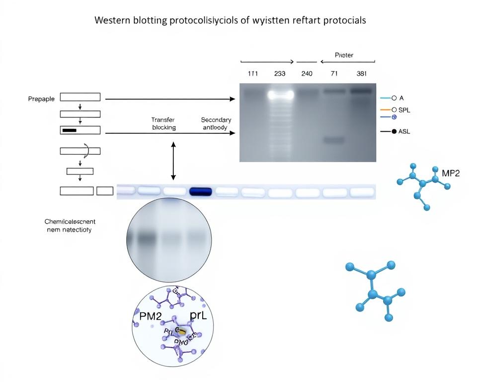
Western blotting, first introduced in 1979, has evolved significantly from a qualitative technique to a powerful quantitative method for protein analysis in modern research. Initially used for detecting the presence or absence of a specific protein in a sample, it now provides a qualitative assessment of changes in protein expression levels. We will explore how this technique has transformed into a precise tool for measuring relative protein abundance.
Understanding how to properly quantify western blot results is essential for generating reliable, reproducible data. This comprehensive guide will walk you through the step-by-step process, from sample preparation to data analysis and interpretation, highlighting the importance of blotting and detection techniques.
Key Takeaways
- Western blotting has evolved into a quantitative method for protein analysis.
- Proper quantification is crucial for reliable and reproducible data.
- Accurate normalization strategies are essential for quantitative western blotting.
- Detection methods play a significant role in the quantification process.
- Practical workflows help generate high-quality, publishable western blot data.
Understanding Western Blot Quantification
The shift from qualitative to quantitative western blotting has significantly impacted various fields of research. Western blot analysis has undergone a significant transformation from its initial qualitative applications to becoming a powerful quantitative tool in modern research laboratories.
The Evolution from Qualitative to Quantitative Analysis
Originally designed to simply detect the presence or absence of specific proteins, western blotting now enables researchers to measure relative changes in protein expression with remarkable precision. The evolution of detection technologies has been crucial in this transition – from film-based detection with limited dynamic range to advanced camera-based systems capable of detecting signals across multiple orders of magnitude.
As noted by experts, “The development of the immunoblot to detect and characterize a protein with an antisera, even in a crude mixture, was a breakthrough with wide-ranging and unpredictable applications across physiology and medicine.”
Why Quantitative Western Blotting Matters in Research
Understanding the fundamental differences between qualitative, semi-quantitative, and fully quantitative western blotting is essential for selecting the appropriate methodology for your research questions. Quantitative western blotting has become increasingly important in fields requiring precise protein measurements, including drug development, disease biomarker identification, and systems biology.
For a detailed guide on western blot protocol, you can refer to our step-by-step guide from sample prep to detection on Western Blot Protocol.
| Application | Qualitative | Quantitative |
|---|---|---|
| Drug Development | Detecting protein presence | Measuring protein expression changes |
| Disease Biomarker Identification | Identifying proteins | Quantifying protein levels |
| Systems Biology | Analyzing protein interactions | Understanding protein regulation |
Proper quantification enables researchers to make meaningful comparisons between experimental conditions, detect subtle changes in protein expression, and generate reproducible data that withstands scientific scrutiny.
Key Requirements for Quantitative Western Blots
For a Western blot to be considered quantitative, it must satisfy specific fundamental requirements. Achieving accurate quantification in Western blotting requires meeting several critical conditions that ensure your signal truly represents the relative abundance of your target protein.
One of the primary conditions is that the signal intensity must be proportional to the amount of protein loaded. This means verifying that the relationship between your protein amount and signal intensity is linear within your working range. To achieve this, you must validate that the signal is not saturated or too low to be detected accurately.
Ensuring Signal Proportionality to Protein Load
Signal proportionality to protein load is fundamental. You must verify that the relationship between your protein amount and signal intensity is linear within your working range. This involves checking that the detection system can accurately measure the changes in protein levels without signal saturation or loss of sensitivity.
Working Within the Linear Range of Detection
Determining the linear range of detection for each target protein is essential, as this range varies significantly between high-abundance proteins (like HSP90) and low-abundance proteins (like Ras). You need to establish the concentration range within which your detection method can accurately quantify the protein of interest.
Implementing Proper Internal Loading Controls
Implementing proper internal loading controls is necessary to normalize for unavoidable variations in sample preparation, gel loading, and transfer efficiency between lanes. Internal loading controls help to ensure that any observed differences in signal intensity are due to true biological changes rather than experimental variability.
| Requirement | Description | Importance |
|---|---|---|
| Signal Proportionality | Signal intensity proportional to protein amount | High |
| Linear Range of Detection | Quantification within the linear range for target proteins | High |
| Internal Loading Controls | Normalization for sample preparation and transfer variability | High |
By meeting these key requirements, you can ensure that your Western blot data is quantitative and reliable, providing valuable insights into the relative abundance of your target proteins.
How to Quantify Western Blot Results Accurately
To achieve precise Western blot quantification, several key factors must be considered. Accurate quantification begins with optimizing the experimental conditions to ensure that the signal detected is proportional to the amount of protein present in the sample.
Optimizing Protein Loading for Quantification
Optimizing protein loading is critical to avoid signal saturation, which occurs when too much protein is loaded onto the gel. We recommend loading smaller amounts of protein (1-10 μg per well) to stay within the linear range of detection. The optimal protein load varies depending on the abundance of the target protein. For instance, high-abundance proteins like HSP90 may require as little as 1-3 μg of total protein, while low-abundance proteins may remain linear up to 40 μg.
To determine the optimal protein load, we suggest creating a standard curve from serial dilutions of a pooled sample representing all experimental conditions. This helps identify the linear range for each target protein, ensuring that the signal is proportional to the protein amount.
Selecting and Diluting Appropriate Antibodies
Antibody dilution optimization is crucial for achieving accurate quantification. Both primary and secondary antibodies should be titrated to find concentrations that provide sufficient sensitivity without causing signal saturation. Typically, primary antibodies are used at dilutions ranging from 1:500 to 1:5,000, while secondary antibodies may require dilutions from 1:50,000 to 1:250,000.
Selecting the right antibodies is essential for specific and sensitive detection of the target protein. We recommend choosing antibodies that have been validated for Western blotting and are known to produce minimal background noise.
Choosing the Right Chemiluminescent Substrate
The choice of chemiluminescent substrate is vital for achieving optimal sensitivity and linear dynamic range. Substrates like SuperSignal West Dura offer a balance between sensitivity and linearity, making them suitable for quantitative applications. Ultrasensitive substrates may cause signal saturation for high-abundance proteins, while standard ECL substrates may lack sufficient sensitivity for low-abundance targets.
Consistent sample preparation, accurate protein concentration determination, and careful gel loading are fundamental prerequisites for reliable quantification. By optimizing these factors and selecting the appropriate reagents, researchers can achieve accurate and meaningful results from their Western blot experiments.

| Protein Abundance | Recommended Protein Load | Typical Antibody Dilution |
|---|---|---|
| High (e.g., HSP90) | 1-3 μg | 1:500 to 1:5,000 |
| Low (e.g., p23) | Up to 40 μg | 1:50,000 to 1:250,000 |
Normalization Strategies for Western Blot Quantification
Normalization is a crucial step in western blot quantification that ensures accurate assessment of target protein abundance. Normalization corrects for unavoidable errors that occur during the western blot process, including sample loading or effects from electrophoresis, transfer, or sample concentration. Choosing the correct normalization method for your quantitative western blot is critical for obtaining reliable and reproducible results.
Housekeeping Proteins as Loading Controls
Traditionally, housekeeping proteins (HKPs) such as β-actin, glyceraldehyde-3-phosphate dehydrogenase (GAPDH), and α-tubulin have been used as loading controls for normalization. However, these proteins can become saturated at common lysate loading amounts, leading to non-linear signals that compromise quantification accuracy. An accurate loading control should display a linear relationship between sample load and signal intensity across all experimental conditions.
Total Protein Normalization (TPN) Methods
Total Protein Normalization (TPN) has emerged as a superior alternative that uses the total protein content in each lane as the normalization factor. TPN methods utilize protein-labeling reagents that covalently attach to proteins, providing a linear response curve with a wide dynamic range for accurate normalization. This approach helps in detecting subtle changes in protein expression and generating reproducible, publishable western blot data.
Comparing Normalization Approaches
When comparing normalization approaches, researchers should consider factors such as linear dynamic range, consistency across experimental conditions, and potential biological regulation of the normalization target. The table below summarizes the key differences between traditional HKP normalization and TPN methods.
| Normalization Method | Characteristics | Advantages |
|---|---|---|
| Housekeeping Proteins (HKPs) | Traditional method using proteins like β-actin, GAPDH | Well-established, easy to implement |
| Total Protein Normalization (TPN) | Uses total protein content in each lane | Wide dynamic range, accurate for detecting subtle changes |
By understanding the strengths and limitations of different normalization strategies, researchers can make informed decisions to improve the accuracy and reliability of their western blot quantification.
Detection Methods: Chemiluminescence vs. Fluorescence
Western blot detection methods have evolved, with chemiluminescence and fluorescence emerging as the two dominant techniques for quantitative analysis. You need to understand the strengths and weaknesses of each method to choose the best approach for your research.
Chemiluminescence Detection: Advantages and Limitations
Chemiluminescence detection uses HRP-conjugated secondary antibodies and substrates to generate light signals that can be captured by camera-based systems or film. The advantages of chemiluminescence include high sensitivity (down to femtogram levels), relatively low cost, and compatibility with existing laboratory equipment. However, it has limitations, including signal saturation, limited multiplexing capabilities, and the production of free radicals that can damage antibodies and membranes.
- High sensitivity down to femtogram levels
- Relatively low cost
- Limited multiplexing capabilities
- Potential for signal saturation
Fluorescence Detection: Multiplexing Capabilities
Fluorescence detection utilizes fluorophore-conjugated secondary antibodies that emit light when excited at specific wavelengths, enabling direct signal measurement. The key advantages of fluorescence include superior multiplexing capabilities (detecting multiple proteins simultaneously), broader linear dynamic range, and elimination of the need for stripping and reprobing membranes. Fluorescence detection is particularly valuable for proteins that resolve at the same molecular weight, as different fluorophores can be used to distinguish between targets.
Selecting the Optimal Detection Method for Your Experiment
When selecting between detection methods, you should consider factors such as target abundance, multiplexing requirements, available equipment, and the need for quantitative precision. By understanding the strengths and limitations of chemiluminescence and fluorescence detection, you can make an informed decision that best suits your experimental needs.
- Consider target abundance and multiplexing requirements
- Assess available equipment and detection method compatibility
- Evaluate the need for quantitative precision
Data Analysis Workflow for Quantitative Western Blots
A robust data analysis workflow is essential for ensuring the reproducibility and accuracy of quantitative western blot results. We will walk you through the critical steps involved in analyzing western blot data, from image acquisition to statistical analysis.
Image Acquisition and Quality Control
Image acquisition begins with capturing high-quality images using camera-based systems that provide a wide dynamic range and avoid signal saturation. You should verify total protein staining patterns, check for consistent transfer efficiency across the membrane, and confirm that signals fall within the linear range of detection.
Densitometric Analysis Techniques
Densitometric analysis involves measuring the intensity of bands using specialized software. This software accurately quantifies signal strength while accounting for background noise. Normalization to loading controls is also crucial to correct for variations in sample loading and transfer efficiency.
Statistical Analysis and Data Interpretation
Statistical analysis should be applied to express results as fold changes relative to control samples, accompanied by measures of variability. Data interpretation requires consideration of both statistical significance and biological relevance, with careful attention to potential technical artifacts.
To illustrate the data analysis workflow, consider the following table that outlines the key components and calculations involved in analyzing western blot data:
| Gel Number | Target | BioGroup_Sample | Band Density |
|---|---|---|---|
| 1 | IBC (Interblot Control) | Pooled Sample | 1000 |
| 1 | LC (Loading Control) | Sample A | 800 |
| 1 | T1 (Target Protein) | Sample A | 1200 |
| 2 | IBC (Interblot Control) | Pooled Sample | 1050 |
| 2 | LC (Loading Control) | Sample B | 850 |
| 2 | T1 (Target Protein) | Sample B | 1300 |
By following this systematic workflow, you can ensure that your western blot data analysis is both accurate and reproducible, providing reliable insights into protein expression differences between samples.
Conclusion
By mastering quantitative Western blotting, researchers can generate high-quality data that advances our understanding of protein biology. Quantitative Western blotting has evolved into a precise technique for measuring relative protein abundance. To achieve this, it’s essential to optimize protein loading, work within the linear range of detection, and implement proper controls. We recommend reducing sample loads to 1-10 μg per well, optimizing antibody dilutions, and using an optimized chemiluminescent substrate. By following these guidelines, you can produce reliable and reproducible Western blot data. As journal standards become more rigorous, mastering quantitative Western blotting is becoming a necessity for researchers publishing protein expression data. With careful attention to technical details, Western blotting can provide valuable insights into protein biology, supporting the development of new therapeutic approaches.
The choice between chemiluminescence and fluorescence detection should be based on experimental requirements. Fluorescence offers superior multiplexing capabilities, while chemiluminescence provides high sensitivity for low-abundance targets. A systematic approach to data analysis, including proper normalization and statistical evaluation, is crucial for extracting meaningful biological insights from Western blot experiments.
References and further readings:
1.Gilda, J. E., & Gomes, A. V. (2013). Western blotting using in-gel protein labeling as a normalization control: stain-free technology. Methods in Molecular Biology, 1295, 381–391.
https://link.springer.com/protocol/10.1007/978-1-4939-2550-6_272.Degasperi, A., Birtwistle, M. R., Volinsky, N., et al. (2014). Evaluating strategies to normalize biological replicates of Western blot data. PLOS ONE, 9(1), e87293.
https://journals.plos.org/plosone/article?id=10.1371/journal.pone.00872933.Taylor, S. C., & Posch, A. (2014). The design of a quantitative western blot experiment. BioMed Research International, 2014, 361590.
https://onlinelibrary.wiley.com/doi/10.1155/2014/361590
FAQ
What are the key considerations for achieving accurate quantification in Western blots?
To achieve accurate quantification, it is essential to ensure signal proportionality to protein load, work within the linear range of detection, and implement proper internal loading controls.
How do I optimize protein loading for quantification in Western blots?
Optimizing protein loading involves loading a consistent amount of protein across all samples, using a reliable method to determine protein concentration, and verifying the integrity of the protein samples.
What is the difference between chemiluminescence and fluorescence detection in Western blots?
Chemiluminescence detection involves the emission of light as a result of a chemical reaction, while fluorescence detection involves the emission of light at a specific wavelength after excitation. Chemiluminescence is often used for sensitive detection, while fluorescence allows for multiplexing capabilities.
What are the advantages of using total protein normalization (TPN) methods?
TPN methods offer several advantages, including reduced variability due to loading errors and the ability to normalize data without relying on a single housekeeping protein.
How do I choose the right chemiluminescent substrate for my Western blot experiment?
The choice of chemiluminescent substrate depends on the sensitivity required, the type of antibody used, and the detection method employed. Selecting a substrate that is compatible with your experimental conditions is crucial.
What are the best practices for densitometric analysis of Western blot data?
Best practices for densitometric analysis include using software that can accurately quantify band intensity, verifying the linearity of the detection system, and normalizing data to internal loading controls.
Can I compare Western blot data across different experiments or gels?
Comparing Western blot data across different experiments or gels requires careful consideration of variables such as protein loading, detection conditions, and normalization methods. Using internal loading controls and normalization strategies can help facilitate comparisons.
Leo Bios
Hello, I’m Leo Bios. As an assistant lecturer, I teach cellular and
molecular biology to undergraduates at a regional US Midwest university. I started as a research tech in
a biotech startup over a decade ago, working on molecular diagnostic tools. This practical experience
fuels my teaching and writing, keeping me engaged in biology’s evolution.


Leave a Comment
Your email address will not be published. Required fields are marked *