Recombinant CD133 Fc-Fused Nanobody (VF0462)
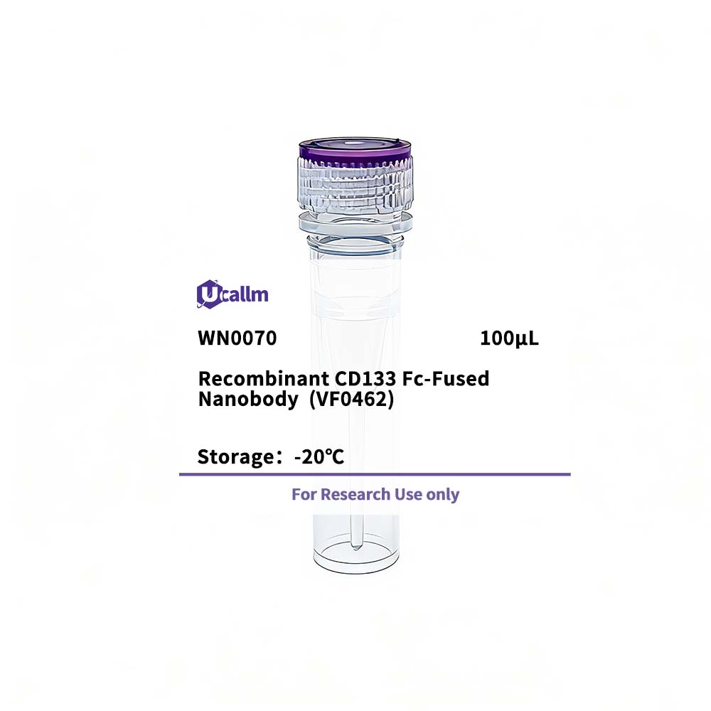
Ucallm_WN0070
$365.00
Product details
Background:This gene encodes a pentaspan transmembrane glycoprotein. The protein localizes to membrane protrusions and is often expressed on adult stem cells, where it is thought to function in maintaining stem cell properties by suppressing differentiation. Mutations in this gene have been shown to result in retinitis pigmentosa and Stargardt disease. Expression of this gene is also associated with several types of cancer. This gene is expressed from at least five alternative promoters that are expressed in a tissue-dependent manner. Multiple transcript variants encoding different isoforms have been found for this gene. [provided by RefSeq, Mar 2009],
Specifications
| Size | 100μL |
| Target | CD133 |
| Reactivity | Human |
| Applications | ELISA |
| MW(Calculated) | |
| MW(Observed) | |
| Host Species | |
| Isotype | IgG2a,Kappa |
| Conjugate/Modification | Unmodified |
| Recommended Dilution Ratio | ELISA 1:5000-100000; Flow Cyt 1-2μg/Test |
| Formulation | Phosphate-buffered solution |
| Purification | Recombinant Expression and Affinity purified |
| Storage | -15°C to -25°C/1 year(Avoid freeze / thaw cycles) |
| Concentration | Please check the information on the tube |
| Clonality | |
| Clone Number | VF0503 |
| Immunogen | Purified recombinant Human CD133 |
| Specificity | This recombinant monoclonal antibody can detects endogenous levels of CD133 protein. |
| Gene Name | PROM1 |
| Protein Name | Prominin-1 |
| Other Name | PROM1; / Prominin-1; / Antigen AC133; / Prominin-like protein 1; / CD133 |
| Species | Human |
| Gene ID | 8842 |
| Uniprot | O43490 |
| Species.1 | |
| Gene ID.1 | |
| Uniprot.1 | |
| Species.2 | |
| Gene ID.2 | |
| Uniprot.2 | |
| Cellular Localization | Apical cell membrane; Multi-pass membrane protein . Cell projection, microvillus membrane; Multi-pass membrane protein . Cell projection, cilium, photoreceptor outer segment . Endoplasmic reticulum. Endoplasmic reticulum-Golgi intermediate compartment. Found in extracellular membrane particles in various body fluids such as cerebrospinal fluid, saliva, seminal fluid and urine. |
| Tissue Expression | Widely distributed with highest levels found in thyroid gland, brain and heart. Expressed in a great number of tumor cells. Expression is down-regulated in different tumors from highly metastatic cells. |
| Signaling_pathway | Human Diseases >> Cancer: overview >> Transcriptional misregulation in cancer |
| Research Areas | |
| Function | Disease:Defects in PROM1 are the cause of cone-rod dystrophy type 12 (CORD12) [MIM:612657]. CORD12 is an inherited retinal dystrophy characterized by retinal pigment deposits visible on fundus examination, predominantly in the macular region, and initial loss of cone photoreceptors followed by rod degeneration. This leads to decreased visual acuity and sensitivity in the central visual field, followed by loss of peripheral vision. Severe loss of vision occurs earlier than in retinitis pigmentosa.,Disease:Defects in PROM1 are the cause of retinal macular dystrophy type 2 (MCDR2) [MIM:608051]. MCDR2 is a bull's-eye macular dystrophy characterized by bilateral annular atrophy of retinal pigment epithelium at the macula.,Disease:Defects in PROM1 are the cause of retinitis pigmentosa type 41 (RP41) [MIM:612095]; also known as retinal degeneration autosomal recessive prominin-related. RP is a retinal dystrophy belonging to the group of pigmentary retinopathies. RP is characterized by retinal pigment deposits visible on fundus examination and primary loss of rod photoreceptor cells followed by secondary loss of cone photoreceptors. Patients typically have night vision blindness and loss of midperipheral visual field. As their condition progresses, they lose their far peripheral visual field and eventually central vision as well.,Disease:Defects in PROM1 are the cause of Stargardt disease type 4 (STGD4) [MIM:603786]. Stargardt disease is the most common hereditary macular degeneration. It is characterized by decreased central vision, atrophy of the macula and underlying retinal pigment epithelium, and frequent presence of prominent flecks in the posterior pole of the retina.,online information:Retina International's Scientific Newsletter,PTM:Glycosylated.,similarity:Belongs to the prominin family.,subunit:Interacts with PCDH21 and with actin filaments.,tissue specificity:Selectively expressed on CD34 hematopoietic stem and progenitor cells in adult and fetal bone marrow, fetal liver, cord blood and adult peripheral blood. Not detected on other blood cells. Also expressed in a number of non-lymphoid tissues including retina, pancreas, placenta, kidney, liver, lung, brain and heart. Found in saliva within small membrane particles., |
Please login to post questions
Related products