SKU: WN0588 Categories: ,

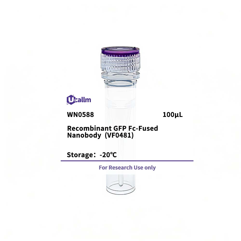
Recombinant GFP Fc-Fused Nanobody (VF0481)

$365.00

SKU: WN0588
Enter the quantity you would like, or drag the panel to find the quantity and price that best for you
Bulk QuoteAuthorize OEM & ODM

Product details

Background:Green Fluorescent Protein (GFP) has quickly become a powerful research tool for assessing gene expression and subcellular protein distribution in fixed or living cells. GFP is excited by and brightly fluoresces when exposed to UV or blue light. This feature makes it ideal as a marker for use in fluorescence microscopy, cytometry, tagging fusion proteins, and assaying transcriptional regulation from gene promoters in vivo. Numerous GFP variants with enhanced and shifted emission spectra (blue, green, and yellow) have been developed through amino acid substitutions at specific residues.
Specifications

Size100μL
TargetGFP
ReactivityHuman
ApplicationsELISA
MW(Calculated)
MW(Observed)
Host Species
IsotypeIgG2a,Kappa
Conjugate/ModificationUnmodified
Recommended Dilution RatioELISA 1:5000-100000; Flow Cyt 1-2μg/Test
FormulationPhosphate-buffered solution
PurificationRecombinant Expression and Affinity purified
Storage-15°C to -25°C/1 year(Avoid freeze / thaw cycles)
ConcentrationPlease check the information on the tube
Clonality
Clone NumberVF0186
ImmunogenPurified recombinant GFP
SpecificityThis recombinant monoclonal antibody can detects endogenous levels of GFP protein.
Gene NameCD4
Protein NameT-cell surface glycoprotein CD4 (T-cell surface antigen T4/Leu-3) (CD antigen CD4)
Other NameGFP nanobody
SpeciesHuman
Gene ID
Uniprot
Species.1
Gene ID.1
Uniprot.1
Species.2
Gene ID.2
Uniprot.2
Cellular LocalizationCell membrane; Single-pass type I membrane protein . Localizes to lipid rafts (PubMed:12517957, PubMed:9168119). Removed from plasma membrane by HIV-1 Nef protein that increases clathrin-dependent endocytosis of this antigen to target it to lysosomal degradation. Cell surface expression is also down-modulated by HIV-1 Envelope polyprotein gp160 that interacts with, and sequesters CD4 in the endoplasmic reticulum.
Tissue ExpressionHighly expressed in T-helper cells. The presence of CD4 is a hallmark of T-helper cells which are specialized in the activation and growth of cytotoxic T-cells, regulation of B cells, or activation of phagocytes. CD4 is also present in other immune cells such as macrophages, dendritic cells or NK cells.
Signaling_pathway
Research Areas
FunctionIntegral membrane glycoprotein that plays an essential role in the immune response and serves multiple functions in responses against both external and internal offenses. In T-cells, functions primarily as a coreceptor for MHC class II molecule:peptide complex. The antigens presented by class II peptides are derived from extracellular proteins while class I peptides are derived from cytosolic proteins. Interacts simultaneously with the T-cell receptor (TCR) and the MHC class II presented by antigen presenting cells (APCs). In turn, recruits the Src kinase LCK to the vicinity of the TCR-CD3 complex. LCK then initiates different intracellular signaling pathways by phosphorylating various substrates ultimately leading to lymphokine production, motility, adhesion and activation of T-helper cells. In other cells such as macrophages or NK cells, plays a role in differentiation/activation, cytokine expression and cell migration in a TCR/LCK-independent pathway. Participates in the development of T-helper cells in the thymus and triggers the differentiation of monocytes into functional mature macrophages.; (Microbial infection) Primary receptor for human immunodeficiency virus-1 (HIV-1) . Down-regulated by HIV-1 Vpu . Acts as a receptor for Human Herpes virus 7/HHV-7 .
Please login to post questions
Back to Top
Product has been added to your cart