SKU: WP14720 Categories: ,

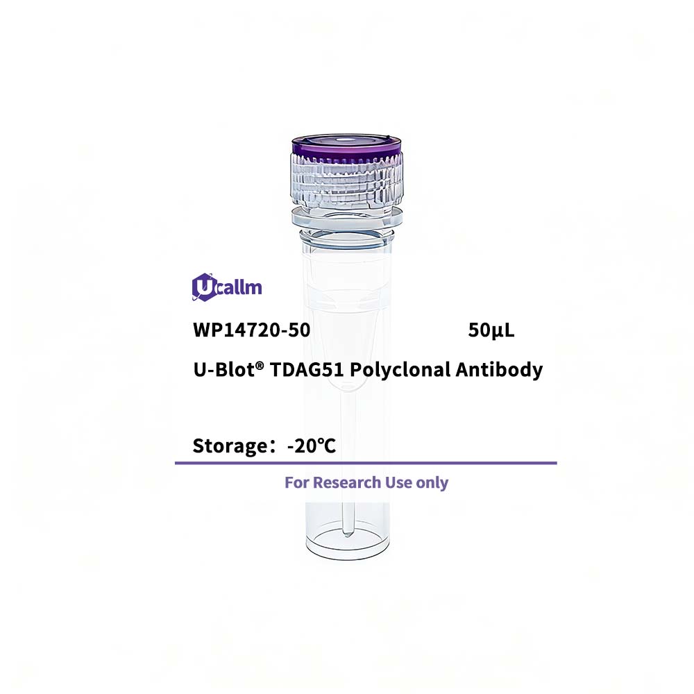
TDAG51 Polyclonal Antibody

Price range: $268.00 through $328.00

SKU: WP14720-100
Enter the quantity you would like, or drag the panel to find the quantity and price that best for you
Bulk QuoteAuthorize OEM & ODM

Product details

Background:

This gene encodes an evolutionarily conserved proline-histidine rich nuclear protein. The encoded protein may play an important role in the anti-apoptotic effects of insulin-like growth factor-1. [provided by RefSeq, Jul 2008],


Specifications

Size50μL/100μL
TargetTDAG51
ReactivityHuman, Mouse, Rat
ApplicationsWB, ELISA
MW(Calculated)
MW(Observed)40kD
Host SpeciesRabbit
IsotypeIgG
Conjugate/ModificationUnmodified
Recommended Dilution RatioWB 1:500-1:2000; ELISA 1:10000; Not yet tested in other applications.
FormulationLiquid in PBS containing 50% glycerol, 0.5% BSA and 0.02% sodium azide.
PurificationThe antibody was affinity-purified from rabbit antiserum by affinity-chromatography using epitope-specific immunogen.
Storage-15°C to -25°C/1 year(Do not lower than -25°C)
Concentration1 mg/ml
ClonalityPolyclonal
Clone Number
ImmunogenThe antiserum was produced against synthesized peptide derived from human PHLA1. AA range:271-320
SpecificityTDAG51 Polyclonal Antibody detects endogenous levels of TDAG51 protein.
Gene NamePHLDA1
Protein NamePleckstrin homology-like domain family A member 1
Other NamePHLDA1; / PHRIP; / TDAG51; / Pleckstrin homology-like domain family A member 1; / Apoptosis-associated nuclear protein; / Proline- and glutamine-rich protein; / PQ-rich protein; / PQR protein; / Proline- and histidine-rich protein; / T-cell death-associated
SpeciesHuman
Gene ID22822
UniprotQ8WV24,
Species.1Mouse
Gene ID.121664
Uniprot.1Q62392,
Species.2Rat
Gene ID.2
Uniprot.2Q9QZA1
Cellular LocalizationCytoplasm . Cytoplasmic vesicle . Nucleus, nucleolus . Colocalizes with intracellular vesicles. .
Tissue ExpressionWidely expressed with highest levels in pancreas. Strongly expressed by benign melanocytic nevi, and progressively reduced expressed in primary and metastatic melanomas (at protein level).
Signaling_pathway
Research Areas
FunctionFunction:Seems to be involved in regulation of apoptosis. May be involved in detachment-mediated programmed cell death. May mediate apoptosis during neuronal development. May be involved in regulation of anti-apoptotic effects of IGF1. May be involved in translational regulation.,induction:Induced by homocysteine and other ER stress-inducing reagents. Induced by phorbolester (TPA)/ionomycin, and stimulation of the T-cell receptor (TCR) complex in T-cells.,similarity:Contains 1 PH domain.,subcellular location:Colocalizes with intracellular vesicles.,subunit:Interacts with RPL14, EIF3S7 and PABPC4.,tissue specificity:Widely expressed with highest levels in pancreas. Strongly expressed by benign melanocytic nevi, and progressively reduced expressed in primary and metastatic melanomas (at protein level).,
Please login to post questions
Back to Top
Product has been added to your cart