

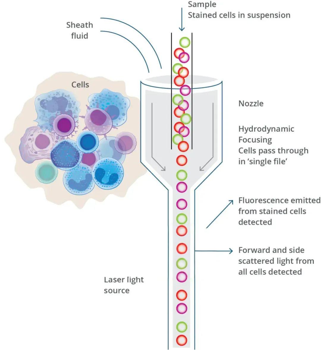
Phosphate-buffered saline (PBS) serves as a fundamental buffer solution in various biological experiments, including flow cytometry. We rely on PBS to maintain cellular integrity and viability during the analysis process. You can trust that PBS provides an isotonic environment, preventing cell damage and ensuring accurate data collection.
PBS is composed of sodium chloride, potassium chloride, sodium phosphate dibasic, and potassium phosphate monobasic, balanced to maintain physiological pH levels. This buffer is crucial for cell suspension and preparation in flow cytometry procedures. By maintaining cell viability, PBS enables researchers to obtain reliable results.
The importance of PBS lies in its ability to create an isotonic environment, making it an essential element in flow cytometry applications. As a result, PBS has become the standard buffer solution for flow cytometry sample preparation and analysis.
Key Takeaways
- PBS is a fundamental buffer solution used in flow cytometry to maintain cellular integrity and viability.
- The composition of PBS includes sodium chloride, potassium chloride, sodium phosphate dibasic, and potassium phosphate monobasic.
- PBS provides an isotonic environment, preventing cell damage during flow cytometry procedures.
- The use of PBS is crucial for obtaining reliable results in flow cytometry experiments.
- PBS has become the standard buffer solution for flow cytometry sample preparation and analysis.
Understanding PBS Solution: Composition and Properties
To comprehend the significance of PBS in flow cytometry, it’s crucial to examine its chemical composition and properties. PBS, or phosphate-buffered saline, is a buffer solution commonly used in biological research.
Chemical Composition of PBS
The chemical composition of PBS includes a precise balance of sodium chloride (NaCl), potassium chloride (KCl), sodium phosphate dibasic (Na2HPO4), and potassium phosphate monobasic (KH2PO4). This balance creates an isotonic environment similar to physiological conditions, making it ideal for maintaining cell viability.
pH and Osmotic Properties
PBS maintains a stable pH of 7.4, closely mimicking the human body’s pH level. The buffering capacity of PBS helps resist pH changes when acids or bases are added, providing a stable environment for cells and biological molecules. Additionally, the osmotic properties of PBS prevent cell shrinkage or swelling during sample preparation and analysis.
Storage and Stability Considerations
Proper storage of PBS is crucial to maintain its effectiveness. Considerations include temperature requirements and potential contamination issues. The stability of PBS under various laboratory conditions is also important, as factors like light exposure and temperature fluctuations can impact its performance in flow cytometry applications.
As we have discussed, the composition and properties of PBS solution are fundamental to its role in flow cytometry. By understanding these aspects, researchers can ensure optimal performance in their experiments.
What Is PBS Solution In Flow Cytometry: Functions and Importance
The use of PBS solution in flow cytometry is essential for maintaining the viability and structural integrity of cells during analysis. PBS, or phosphate-buffered saline, serves as a critical component in the preparation and analysis of cells for flow cytometry.
Role as a Cell Suspension Buffer
PBS solution acts as the primary cell suspension buffer in flow cytometry, providing an optimal environment for cells to be analyzed. Its isotonic nature prevents osmotic stress on cells, which is crucial for accurate measurements as cell size and granularity are key parameters in flow cytometry. By maintaining cellular integrity, PBS ensures that the results obtained from flow cytometry are reliable and accurate.
Maintaining Cell Viability During Analysis
One of the critical functions of PBS solution in flow cytometry is to maintain cell viability during the analysis process. The sometimes lengthy preparation and analysis procedures can be detrimental to cell health, but PBS helps to mitigate this issue by providing a gentle and non-toxic environment. This is particularly important in flow cytometry, where cell viability can significantly impact the accuracy of the results.
Compatibility with Antibodies and Fluorochromes
PBS solution is also notable for its compatibility with various antibodies and fluorochromes used in flow cytometry. This compatibility makes PBS an ideal buffer for immunophenotyping and other fluorescence-based detection methods. By minimizing background fluorescence and non-specific binding, PBS solution enhances the sensitivity of detection and improves signal-to-noise ratios in flow cytometry analysis.
| Key Functions of PBS in Flow Cytometry | Description |
|---|---|
| Cell Suspension Buffer | Provides an optimal environment for cell analysis, preventing osmotic stress. |
| Maintaining Cell Viability | Ensures cell health during preparation and analysis, improving result accuracy. |
| Compatibility with Antibodies and Fluorochromes | Enhances sensitivity and reduces background fluorescence in flow cytometry. |
In conclusion, PBS solution plays a multifaceted role in flow cytometry, serving not only as a cell suspension buffer but also in maintaining cell viability and ensuring compatibility with various detection methods. Its importance cannot be overstated, as it directly impacts the accuracy and reliability of flow cytometry results.

Applications of PBS in Flow Cytometry Protocols
The application of PBS in flow cytometry is multifaceted, encompassing cell preparation, washing, and staining protocols that are essential for accurate cellular analysis. PBS serves as a foundational buffer in these processes, ensuring the integrity and viability of cells during various experimental procedures.
Cell Preparation and Washing Procedures
PBS is integral to cell preparation procedures in flow cytometry, serving as the primary washing buffer for removing unwanted debris, media components, and unbound reagents from cell samples. To prepare cells, we typically use PBS/BSA buffer (phosphate-buffered saline pH 7.4 and 1% BSA). For tissue culture cell lines in suspension, cells are decanted into 15 ml conical centrifuge tubes, centrifuged at 400 g for 5 minutes, and then resuspended in 10 ml of PBS/BSA.
| Step | Procedure | Reagent |
|---|---|---|
| 1 | Decant cells into 15 ml conical centrifuge tube | Tissue culture cell lines |
| 2 | Centrifuge at 400 g for 5 minutes | PBS/BSA buffer |
| 3 | Resuspend pellet in 10 ml of PBS/BSA | PBS/BSA buffer |
Antibody Staining Protocols
For direct immunofluorescence staining, cells are prepared and adjusted to a concentration of 1 × 10^6 cells/ml with PBS/BSA buffer. We aliquot 100 μl of cell suspension into test tubes, add antibody at the recommended dilution, and incubate at room temperature for 30 minutes. After staining, cells are washed with 2 ml of PBS/BSA, centrifuged at 400 g for 5 minutes, and the supernatant is discarded.
Intracellular Staining Applications
Intracellular staining applications require specialized PBS-based buffers for cell permeabilization and fixation. We use these buffers to facilitate the entry of antibodies into cells, allowing for the staining of intracellular antigens. The process involves fixing cells with a fixative, permeabilizing with a detergent, and then staining with antibodies diluted in PBS/BSA.
PBS Modifications for Specialized Applications
PBS can be modified for specialized applications by adding reagents such as BSA, sodium azide, or EDTA. These modifications enhance the performance of PBS in specific flow cytometry protocols. For example, adding BSA helps to block non-specific binding sites, while sodium azide prevents microbial growth.
By understanding the various applications and modifications of PBS in flow cytometry protocols, researchers can optimize their experimental procedures to achieve more accurate and reliable results.
Conclusion: Best Practices for Using PBS in Flow Cytometry
Ultimately, the use of PBS solution in flow cytometry is essential for maintaining cell viability and ensuring the integrity of experimental results. PBS serves as an ideal vehicle for diluting reagents, washing cells, and performing biological assays due to its compatibility with a wide range of biological molecules.
To optimize PBS usage in flow cytometry, researchers should adhere to best practices for preparing, storing, and using PBS. This includes quality control measures to ensure buffer integrity and avoiding common mistakes such as improper storage or contamination.
Standardization and Optimization: Standardizing PBS preparation and usage across laboratories is crucial for ensuring comparable and reproducible flow cytometry results. Researchers should consider optimizing PBS formulations for specific research applications, including adjustments for different cell types and experimental conditions.
Future Trends: As flow cytometry continues to evolve, the development of specialized buffer formulations will play a critical role in advancing research capabilities. By understanding the importance of PBS solution in flow cytometry and implementing best practices, researchers can ensure reliable and reproducible results in their studies.
References and further readings:
1.Riccardi, C., & Nicoletti, I. (2006). Analysis of apoptosis by propidium iodide staining and flow cytometry. Nature Protocols, 1(3), 1458–1461.
https://www.nature.com/articles/nprot.2006.2382.Perez, O. D., Krutzik, P. O., & Nolan, G. P. (2004). Flow cytometric analysis of kinase signaling cascades. Methods in Molecular Biology, 263, 67–94.
https://link.springer.com/protocol/10.1385/1-59259-773-4:0673.Foster, B., Prussin, C., Liu, F., & Whitmire, J. K. (2007). Detection of intracellular cytokines by flow cytometry. Current Protocols in Immunology, 6.24.
https://currentprotocols.onlinelibrary.wiley.com/doi/10.1002/0471142735.im0624s78
FAQ
What is the ideal pH range for PBS used in flow cytometry?
The ideal pH range for PBS is between 7.2 and 7.4, which is isotonic and non-toxic to most cells, maintaining their viability during analysis.
Can PBS be used as a substitute for other buffers in flow cytometry protocols?
PBS is a versatile buffer, but its suitability depends on the specific requirements of the protocol. For certain applications, other buffers may be more appropriate due to differences in ionic strength or the presence of specific ions like magnesium or calcium.
How should PBS be stored to maintain its stability?
PBS should be stored at room temperature, away from direct light, and in a tightly sealed container to prevent contamination and degradation. It’s also recommended to check the buffer’s pH and sterility before use.
Is it necessary to filter PBS before using it in flow cytometry?
Yes, filtering PBS through a 0.2-micron filter is recommended to remove any particulate matter or contaminants that could interfere with flow cytometry analysis or clog the instrument.
Can PBS be used for washing cells before antibody staining?
Yes, PBS is commonly used for washing cells to remove residual culture media or other substances that might interfere with antibody staining or flow cytometry analysis.
Are there any modifications to PBS that can enhance its performance in specific flow cytometry applications?
Yes, modifications such as adding bovine serum albumin (BSA) or fetal bovine serum (FBS) can help reduce non-specific binding of antibodies, while adding azide can help prevent microbial growth. The specific modification depends on the application and experimental requirements.
What are the consequences of using PBS that has degraded or is out of the optimal pH range?
Using degraded or pH-compromised PBS can lead to reduced cell viability, altered cell surface antigen expression, or increased non-specific staining, ultimately affecting the accuracy and reliability of flow cytometry results.
Leo Bios
Hello, I’m Leo Bios. As an assistant lecturer, I teach cellular and
molecular biology to undergraduates at a regional US Midwest university. I started as a research tech in
a biotech startup over a decade ago, working on molecular diagnostic tools. This practical experience
fuels my teaching and writing, keeping me engaged in biology’s evolution.


Leave a Comment
Your email address will not be published. Required fields are marked *