SKU: WM1234 Categories: ,

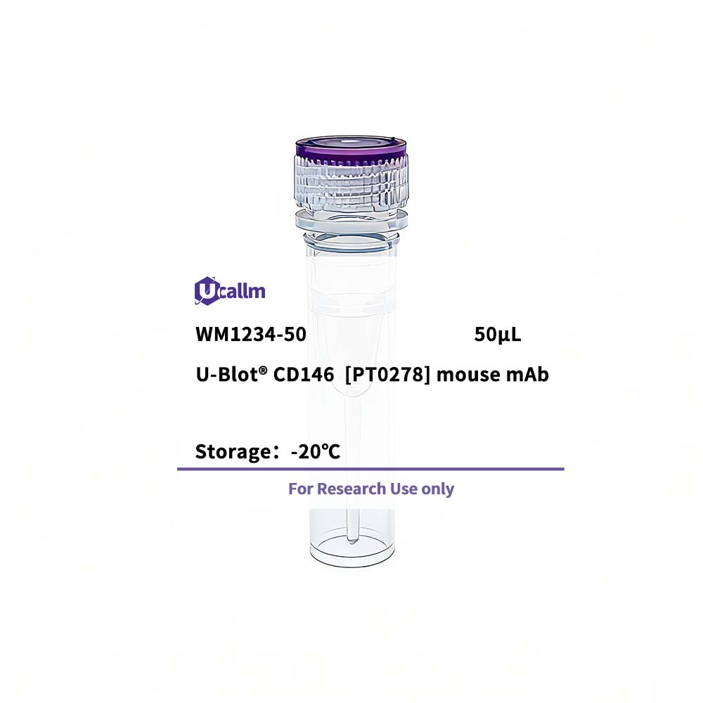
CD146 [PT0278] mouse mAb

Price range: $268.00 through $328.00

SKU: WM1234-50
Enter the quantity you would like, or drag the panel to find the quantity and price that best for you
Bulk QuoteAuthorize OEM & ODM

Product details

Background:
function:Plays a role in cell adhesion, and in cohesion of the endothelial monolayer at intercellular junctions in vascular tissue. Its expression may allow melanoma cells to interact with cellular elements of the vascular system, thereby enhancing hematogeneous tumor spread. Could be an adhesion molecule active in neural crest cells during embryonic development. Acts as surface receptor that triggers tyrosine phosphorylation of FYN and PTK2, and a transient increase in the intracellular calcium concentration., similarity:Contains 2 Ig-like V-type (immunoglobulin-like) domains., similarity:Contains 3 Ig-like C2-type (immunoglobulin-like) domains., tissue specificity:Detected in endothelial cells in vascular tissue throughout the body. May appear at the surface of neural crest cells during their embryonic migration. Appears to be limited to vascular smooth muscle in normal adult tissues. Associated with tumor progression and the development of metastasis in human malignant melanoma. Expressed most strongly on metastatic lesions and advanced primary tumors and is only rarely detected in benign melanocytic nevi and thin primary melanomas with a low probability of metastasis.,

Specifications

Size50μL/100μL
TargetCD146
ReactivityHuman
ApplicationsIHC, WB, IF, ELISA
MW(Calculated)72kD
MW(Observed)110kD
Host SpeciesMouse
IsotypeIgG1,Kappa
Conjugate/ModificationUnmodified
Recommended Dilution RatioIHC 1:200-1000; WB 1:500-2000; IF 1:100-500; ELISA 1:1000-5000 / Note: For IHC, we suggest antigen retrieval with TE buffer pH 9.0 (Cat#WT1004)
FormulationPBS, 50% glycerol, 0.05% Proclin 300, 0.05%BSA
PurificationRecombinant Antibody expressed in animal component-free (ACF) media, purified via Protein G affinity chromatography.
Storage-15°C to -25°C/1 year(Do not lower than -25°C)
Concentration
ClonalityMonoclonal
Clone NumberRMM0278
ImmunogenSynthesized peptide derived from human CD146 AA range: 500-600
SpecificityThis antibody detects endogenous levels of CD146 protein.
Gene NameMCAM MUC18
Protein NameCD146
Other NameCell surface glycoprotein MUC18; / Cell surface glycoprotein P1H12; / Melanoma cell adhesion molecule; / Melanoma-associated antigen A32; / Melanoma-associated antigen MUC18; / S-endo 1 endothelial-associated antigen; / CD antigen CD146
SpeciesHuman
Gene ID4162
UniprotP43121
Species.1
Gene ID.1
Uniprot.1
Species.2
Gene ID.2
Uniprot.2
Cellular LocalizationMembranous, Cytoplasmic
Tissue ExpressionDetected in endothelial cells in vascular tissue throughout the body. May appear at the surface of neural crest cells during their embryonic migration. Appears to be limited to vascular smooth muscle in normal adult tissues. Associated with tumor progression and the development of metastasis in human malignant melanoma. Expressed most strongly on metastatic lesions and advanced primary tumors and is only rarely detected in benign melanocytic nevi and thin primary melanomas with a low probability of metastasis.
Signaling_pathway
Research Areas
FunctionFunction:Plays a role in cell adhesion, and in cohesion of the endothelial monolayer at intercellular junctions in vascular tissue. Its expression may allow melanoma cells to interact with cellular elements of the vascular system, thereby enhancing hematogeneous tumor spread. Could be an adhesion molecule active in neural crest cells during embryonic development. Acts as surface receptor that triggers tyrosine phosphorylation of FYN and PTK2, and a transient increase in the intracellular calcium concentration.,similarity:Contains 2 Ig-like V-type (immunoglobulin-like) domains.,similarity:Contains 3 Ig-like C2-type (immunoglobulin-like) domains.,tissue specificity:Detected in endothelial cells in vascular tissue throughout the body. May appear at the surface of neural crest cells during their embryonic migration. Appears to be limited to vascular smooth muscle in normal adult tissues. Associated with tumor progression and the development of metastasis in human malignant melanoma. Expressed most strongly on metastatic lesions and advanced primary tumors and is only rarely detected in benign melanocytic nevi and thin primary melanomas with a low probability of metastasis.,
Please login to post questions
Back to Top
Product has been added to your cart