Product details
Background:
tissue specificity:Expression is restricted to melanoma and melanocyte cell lines and retina.,
Price range: $268.00 through $328.00
Background:
tissue specificity:Expression is restricted to melanoma and melanocyte cell lines and retina.,
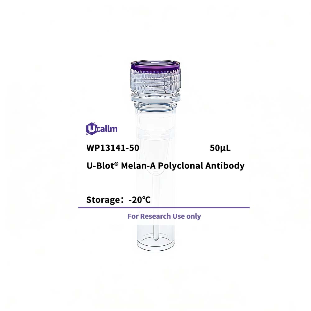
| Size | 50μL/100μL |
| Target | Melan-A |
| Reactivity | Human |
| Applications | WB, IHC, IF, ELISA |
| MW(Calculated) | |
| MW(Observed) | 15kD |
| Host Species | Rabbit |
| Isotype | IgG |
| Conjugate/Modification | Unmodified |
| Recommended Dilution Ratio | WB 1:500-1:2000; IHC 1:100-1:300; ELISA 1:40000; IF 1:50-200 / Note: For IHC, we suggest antigen retrieval with TE buffer pH 9.0 (Cat#WT1004) |
| Formulation | Liquid in PBS containing 50% glycerol, 0.5% BSA and 0.02% sodium azide. |
| Purification | The antibody was affinity-purified from rabbit antiserum by affinity-chromatography using epitope-specific immunogen. |
| Storage | -15°C to -25°C/1 year(Do not lower than -25°C) |
| Concentration | 1 mg/ml |
| Clonality | Polyclonal |
| Clone Number | |
| Immunogen | The antiserum was produced against synthesized peptide derived from human MART-1. AA range:41-90 |
| Specificity | Melan-A Polyclonal Antibody detects endogenous levels of Melan-A protein. |
| Gene Name | MLANA |
| Protein Name | Melanoma antigen recognized by T-cells 1 |
| Other Name | MLANA; / MART1; / Melanoma antigen recognized by T-cells 1; / MART-1; / Antigen LB39-AA; / Antigen SK29-AA; / Protein Melan-A |
| Species | Human |
| Gene ID | 2315 |
| Uniprot | Q16655 |
| Species.1 | |
| Gene ID.1 | |
| Uniprot.1 | |
| Species.2 | |
| Gene ID.2 | |
| Uniprot.2 | |
| Cellular Localization | Endoplasmic reticulum membrane; Single-pass type III membrane protein. Golgi apparatus. Golgi apparatus, trans-Golgi network membrane. Melanosome. Also found in small vesicles and tubules dispersed over the entire cytoplasm. A small fraction of the protein is inserted into the membrane in an inverted orientation. Inversion of membrane topology results in the relocalization of the protein from a predominant Golgi/post-Golgi area to the endoplasmic reticulum. Melanoma cells expressing the protein with an inverted membrane topology are more effectively recognized by specific cytolytic T-lymphocytes than those expressing the protein in its native membrane orientation. |
| Tissue Expression | Expression is restricted to melanoma and melanocyte cell lines and retina. |
| Signaling_pathway | |
| Research Areas | |
| Function | tissue specificity:Expression is restricted to melanoma and melanocyte cell lines and retina., |