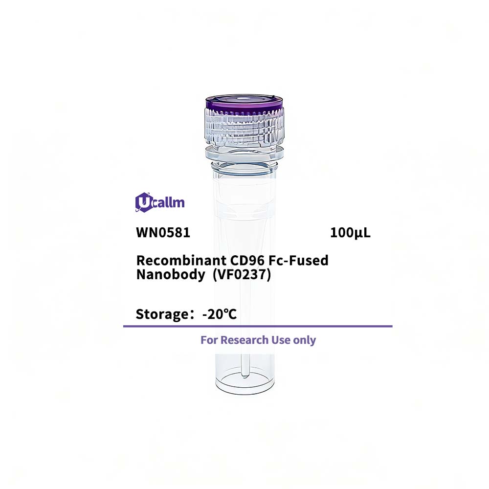
SKU: WN0581 Categories: ,

Recombinant CD96 Fc-Fused Nanobody (VF0237)

$365.00

SKU: WN0581
Enter the quantity you would like, or drag the panel to find the quantity and price that best for you
Bulk QuoteAuthorize OEM & ODM

Product details

Background:The protein encoded byThis gene belongs to the immunoglobulin superfamily. It is a type I membrane protein. The protein may play a role in the adhesive interactions of activated T and NK cells during the late phase of the immune response. It may also function in antigen presentation. Alternative splicing generates multiple transcript variants encoding distinct isoforms. [provided by RefSeq, Jan 2016]

Specifications

Size100μL
TargetCD96
ReactivityHuman
ApplicationsELISA
MW(Calculated)
MW(Observed)
Host Species
IsotypeIgG2a,Kappa
Conjugate/ModificationUnmodified
Recommended Dilution RatioELISA 1:5000-100000
FormulationPhosphate-buffered solution
PurificationRecombinant Expression and Affinity purified
Storage-15°C to -25°C/1 year(Avoid freeze / thaw cycles)
ConcentrationPlease check the information on the tube
Clonality
Clone NumberVF0658
ImmunogenPurified recombinant Human CD96
SpecificityThis recombinant monoclonal antibody can detects endogenous levels of CD96 protein.
Gene NameCD96
Protein NameT-cell surface protein tactile (Cell surface antigen CD96) (T cell-activated increased late expression protein) (CD antigen CD96)
Other NameCD96; / T-cell surface protein tactile; / Cell surface antigen CD96; / T cell-activated increased late expression protein; / CD96; / CD96 nanobody
SpeciesHuman
Gene ID10225
UniprotP40200
Species.1
Gene ID.1
Uniprot.1
Species.2
Gene ID.2
Uniprot.2
Cellular LocalizationMembrane; Single-pass type I membrane protein.
Tissue ExpressionDetected on hematopoietic stem cells derived from umbilical cord blood (PubMed:2474813). Detected on lymph vessel endothelial cells, skin and tonsil (PubMed:23169771). Detected on peripheral blood monocytes (PubMed:154873). Detected on monocyte-derived dendritic cells (at protein level) (PubMed:1635286). Detected at low levels in spleen, placenta, liver (PubMed:952422). Expressed by activated T-cells, B-cells, monocytes and thymic epithelial cells (PubMed:7767). Isoform 1 and isoform 3 are detected in vein and artery endothelial cells, astrocytes, keratinocytes and artery smooth muscle cells (PubMed:15496415). Expressed by neurons in the brain. Restricted expression in tumor cell lines. Detected in highly metastasizing melanoma cell lines (PubMed:952422).
Signaling_pathway
Research Areas
Functiondevelopmental stage:Expressed at low levels on peripheral T-cells and is strongly up-regulated after activation, peaking 6 to 9 days after the activating stimulus.,Disease:A chromosomal aberration involving CD96 is associated with C syndrome [MIM:211750]. Translocation t(3; 18)(q13.13; q12.1). CD96 gene was located at the 3q13.13 breakpoint. Precise structural analysis around the breakpoint showed that the gene was disrupted by the translocation in exon 5, probably leading to premature termination or loss of expression of CD96 protein. No gene was detected at the chromosome 18 breakpoint.,Disease:Defects in CD96 are a cause of C syndrome [MIM:211750]; also called Opitz trigonocephaly syndrome. This syndrome is characterized by trigonocephaly and associated anomalies, such as unusual facies, wide alveolar ridges, multiple buccal frenula, limb defects, visceral anomalies, redundant skin, psychomotor retardation and hypotonia.,Disease:Defects in CD96 are a cause of C-like syndrome [MIM:605039]; also called Opitz trigonocephaly-like syndrome. The C-like syndrome seems to be a severe form of the C syndrome. It is controversial whether there is (1) a gradient of spectrum in the C syndrome, from the mild form (C syndrome) to the severe form (C-like syndrome), or (2) genetic heterogeneity among the patients with the C syndrome.,May be involved in adhesive interactions of activated T and NK cells during the late phase of the immune response. Promotes NK cell-target adhesion by interacting with PVR present on target cells. May function at a time after T and NK cells have penetrated the endothelium using integrins and selectins, when they are actively engaging diseased cells and moving within areas of inflammation.,similarity:Contains 1 Ig-like C2-type (immunoglobulin-like) domain.,similarity:Contains 2 Ig-like V-type (immunoglobulin-like) domains.,subunit:Homodimer; disulfide-linked. Interacts with PVR.,tissue specificity:Expressed on normal T-cell lines and clones, and some transformed T-cells, but no other cultured cell lines tested. It is expressed at very low levels on activated B-cells.,
Please login to post questions
Back to Top
Product has been added to your cart