U-Blot® Villin Rabbit mAb (Ready to Use)
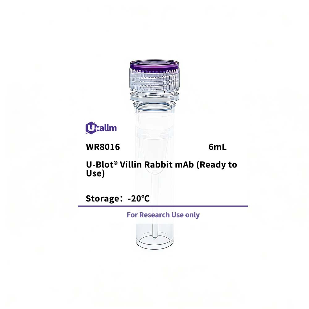
Ucallm_WR8016
$168.00
Product details
Background:
This gene encodes a member of a family of calcium-regulated actin-binding proteins. This protein represents a dominant part of the brush border cytoskeleton which functions in the capping, severing, and bundling of actin filaments. Two mRNAs of 2.7 kb and 3.5 kb have been observed; they result from utilization of alternate poly-adenylation signals present in the terminal exon. [provided by RefSeq, Jul 2008],
Specifications
| Size | 6mL |
| Target | Villin |
| Reactivity | Human, Mouse, Pig, Bovine |
| Applications | IHC |
| MW(Calculated) | |
| MW(Observed) | |
| Host Species | Rabbit |
| Isotype | IgG1,Kappa |
| Conjugate/Modification | Unmodified |
| Recommended Dilution Ratio | Ready to use for IHC / Note: For IHC, we suggest antigen retrieval with TE buffer pH 9.0 (Cat#WT1004) |
| Formulation | The prediluted ready-to-use antibody is diluted in phosphate buffer saline containing stabilizing protein and 0.05% Proclin 300 |
| Purification | Recombinant Expression and Affinity purified |
| Storage | 2°C to 8°C/1 year,Ship by ice bag |
| Concentration | |
| Clonality | Monoclonal |
| Clone Number | RM0094R |
| Immunogen | Synthesized peptide derived from human Villin AA range:150-250 |
| Specificity | This antibody detects endogenous levels of Villin |
| Gene Name | VIL1 VIL |
| Protein Name | Villin-1 |
| Other Name | D2S1471; / OTTHUMP00000164145; / VIL; / VIL1; / VILI_HUMAN; / Villin 1; / Villin-1; / Villin1 |
| Species | Human |
| Gene ID | 7429 |
| Uniprot | P09327 |
| Species.1 | |
| Gene ID.1 | |
| Uniprot.1 | |
| Species.2 | |
| Gene ID.2 | |
| Uniprot.2 | |
| Cellular Localization | Cytoplasm, cytoskeleton. Cell projection, lamellipodium. Cell projection, ruffle. Cell projection, microvillus. Cell projection, filopodium tip . Cell projection, filopodium . Relocalized in the tip of cellular protrusions and filipodial extensions upon infection with S.flexneri in primary intestinal epithelial cells (IEC) and in the tail-like structures forming the actin comets of S.flexneri. Redistributed to the leading edge of hepatocyte growth factor (HGF)-induced lamellipodia (By similarity). Rapidly redistributed to ruffles and lamellipodia structures in response to autotaxin, lysophosphatidic acid (LPA) and epidermal growth factor (EGF) treatment. . |
| Tissue Expression | Specifically expressed in epithelial cells. Major component of microvilli of intestinal epithelial cells and kidney proximal tubule cells. Expressed in canalicular microvilli of hepatocytes (at protein level). |
| Signaling_pathway | |
| Research Areas | |
| Function | Domain:Consists of a large core fragment, the N-terminal portion, and a small headpiece, the C-terminal portion. The headpiece binds F-actin strongly in both the presence and absence of calcium.,Function:Ca(2+)-regulated actin-binding protein.,similarity:Belongs to the villin/gelsolin family.,similarity:Contains 1 HP (headpiece) domain.,similarity:Contains 6 gelsolin-like repeats.,subunit:Monomer.,tissue specificity:Major component of microvilli of intestinal epithelial cells and kidney proximal tubule cells., |
Please login to post questions
Related products