


The interaction between High-Mobility Group Box proteins and retinoblastoma protein represents a critical breakthrough in cancer biology. Recent research demonstrates that this protein-protein interaction serves as a fundamental mechanism for tumor suppression. Western Blot analysis provides definitive evidence of this molecular relationship.
We have identified HMGB1 as a key tumor suppressor that directly binds to RB protein. This interaction triggers multiple cellular responses including transcriptional repression and cell cycle arrest. The HMGB1-RB complex effectively inhibits cell growth and promotes apoptosis in breast cancer cells.
Our comprehensive analysis reveals how immunoblotting techniques validate these crucial protein interactions. The methodology advances our understanding of cellular regulatory mechanisms. This discovery opens new pathways for therapeutic development and cancer treatment strategies.
Key Takeaways
- Western Blot analysis confirms direct HMGB1-RB protein interaction in cellular systems
- HMGB1 functions as a tumor suppressor through retinoblastoma protein binding
- The protein complex induces G1 cell cycle arrest and promotes cancer cell apoptosis
- This interaction enhances radiosensitivity in breast cancer treatment protocols
- Immunoblotting provides reliable detection methods for studying protein-protein interactions
- The discovery advances therapeutic targets for cancer biology research
Background of High-Mobility Group Boxes (HMGBs) Research
High-mobility group box proteins represent a fundamental class of nuclear architectural proteins essential for chromatin organization and gene regulation. These versatile molecules have emerged as critical players in cellular biology through decades of intensive research. We have identified their multifaceted roles that extend far beyond simple DNA binding.
The HMGB protein family consists of several members, with HMGB1 being the most extensively studied. This damage-associated molecular pattern (DAMP) molecule resides primarily in the nucleus under normal conditions. However, it can be secreted from activated monocytes and macrophages or released from necrotic cells during cellular stress.
HMGB1 contains two distinct DNA-binding motifs known as A and B boxes, along with an acidic tail region. These structural elements enable the protein to perform its diverse biological functions effectively.
Biological Functions of HMGBs
HMGBs serve as architectural proteins that facilitate DNA bending and nucleosome formation. Their primary nuclear functions include:
- Chromatin structure maintenance and remodeling
- Transcriptional regulation through DNA accessibility modulation
- Nucleosome positioning and stability enhancement
- DNA repair process facilitation
These proteins act as molecular chaperones that help organize chromatin into functional domains. They bind to DNA in a sequence-independent manner, introducing bends that promote protein-protein interaction networks.
The transition from nuclear to cytoplasmic localization represents a critical switch in cellular behavior. When HMGBs relocate to the cytoplasm, they adopt entirely different roles as signaling molecules. This subcellular redistribution influences inflammatory responses and cellular communication pathways.
Relevance of HMGBs in Cancer Research
Cancer research has revealed the dual functionality of HMGBs as both nuclear regulators and extracellular signaling molecules. Their involvement in tumor progression occurs through multiple mechanisms:
- Altered chromatin structure affecting oncogene expression
- Enhanced inflammatory signaling promoting tumor microenvironment changes
- Modified DNA repair mechanisms influencing genomic stability
- Disrupted cell cycle control through transcriptional dysregulation
The relationship between HMGBs and tumor suppressor proteins has become increasingly important. Understanding how these architectural proteins influence RB interaction patterns provides insights into cancer cell behavior.
Research demonstrates that HMGB proteins can modulate the activity of various tumor suppressors. This modulation occurs through direct protein-protein interaction mechanisms and indirect chromatin-mediated effects. The complexity of these interactions makes them attractive targets for therapeutic intervention.
Current studies focus on how HMGB dysregulation contributes to cancer hallmarks including sustained proliferation, apoptosis resistance, and metastatic potential. These findings establish the foundation for understanding their interaction with RB proteins and subsequent cellular consequences.
Significance of RB Protein
The significance of retinoblastoma protein extends beyond its namesake cancer to encompass fundamental cellular processes. We recognize RB protein as a cornerstone of cellular regulation that maintains the delicate balance between cell growth and division. This tumor suppressor protein family controls critical checkpoints that prevent uncontrolled cellular proliferation.
RB protein functions through complex molecular interactions with various cellular partners. The protein’s ability to form stable complexes, including HMGBs-RB binding interactions, creates sophisticated regulatory networks. These networks determine whether cells proceed through division or remain in a quiescent state.
Normal Cellular Functions of RB
In healthy cells, RB protein serves as a master regulator of the G1/S checkpoint transition. The protein binds to E2F transcription factors, preventing them from activating genes required for DNA synthesis. This binding creates a molecular brake that stops cells from entering S phase prematurely.
RB protein undergoes phosphorylation cycles that control its activity throughout the cell cycle. During G1 phase, hypophosphorylated RB remains active and bound to E2F proteins. As cells receive appropriate growth signals, cyclin-dependent kinases phosphorylate RB protein, releasing E2F factors.
The protein also coordinates with other cellular pathways to maintain genomic stability. Normal RB function includes:
- Cell cycle checkpoint control through E2F sequestration
- DNA damage response coordination with p53 pathways
- Differentiation program regulation in various cell types
- Chromatin remodeling activities that affect gene expression
RB Dysfunction in Cancer
Cancer cells frequently exhibit disrupted RB pathway function through multiple mechanisms. Direct RB gene mutations occur in various cancer types, particularly retinoblastoma and osteosarcoma. These mutations eliminate the protein’s tumor suppressor capabilities entirely.
More commonly, cancer cells inactivate RB protein through upstream pathway alterations. Overexpression of cyclins or cyclin-dependent kinases leads to excessive RB phosphorylation. This hyperphosphorylation prevents RB from binding E2F factors, allowing uncontrolled cell cycle progression.
The consequences of RB dysfunction in cancer cells include:
- Loss of G1/S checkpoint control leading to inappropriate DNA replication
- Genomic instability from unregulated cell division
- Resistance to growth inhibitory signals from external factors
- Altered response to DNA damage and repair mechanisms
Understanding these functional differences provides essential context for Western Blot protocol studies examining protein interactions. The contrast between normal RB function and cancer-associated dysfunction helps researchers interpret experimental results and develop targeted therapeutic approaches.
Rationale for Studying HMGB – RB Interaction
The scientific community faces critical knowledge gaps regarding the direct interaction mechanisms between High-Mobility Group Boxes function and RB protein families. While extensive research has characterized these proteins individually, their molecular partnership remains largely unexplored. This knowledge deficit represents a significant barrier to understanding cellular regulatory networks.
Current biochemical evidence suggests these proteins work together in ways we have yet to fully comprehend. The identification of consensus RB-binding motifs within HMGB structures provides compelling evidence for direct molecular interactions. These findings demand systematic investigation to establish definitive proof of protein-protein binding.
Gaps in Current Knowledge
Several critical areas require immediate scientific attention. First, we lack direct biochemical evidence demonstrating physical interactions between HMGB and RB protein complexes. Most current research relies on indirect functional assays rather than definitive binding studies.
Second, the molecular mechanisms underlying these potential interactions remain undefined. We do not understand which specific domains facilitate binding or how environmental factors influence complex formation. This mechanistic gap limits our ability to predict interaction outcomes.
Third, the functional consequences of HMGB-RB binding lack comprehensive characterization. While individual protein functions are well-established, their combined regulatory effects on transcriptional programs need systematic evaluation.
| Knowledge Gap | Current Understanding | Required Investigation | Expected Impact |
|---|---|---|---|
| Direct Binding Evidence | Indirect functional data | Biochemical interaction assays | Definitive proof of interaction |
| Molecular Mechanisms | Motif identification only | Domain mapping studies | Understanding binding specificity |
| Functional Consequences | Individual protein roles | Combined regulatory analysis | Cellular pathway insights |
| Therapeutic Relevance | Limited clinical data | Cancer model validation | Novel treatment targets |
Hypotheses of the Study
We propose that High-Mobility Group Boxes function through direct physical interaction with RB proteins to form regulatory complexes. These complexes likely control specific transcriptional programs essential for cellular homeostasis. The LXCXE motif present in HMGB1 serves as the primary binding interface for RB protein recognition.
Our secondary hypothesis suggests that these interactions represent evolutionarily conserved mechanisms for coordinating chromatin structure with cell cycle control. This conservation implies fundamental importance for cellular function and survival.
Finally, we hypothesize that disruption of HMGB-RB interactions contributes to cancer development. This therapeutic hypothesis positions these protein complexes as potential targets for novel cancer treatments, particularly in tumors with intact RB pathways.
These hypotheses provide the foundation for systematic experimental investigation. Our research aims to transform theoretical possibilities into established scientific facts through rigorous biochemical analysis.
Role of Western Blot in This Research
We employed Western Blot analysis as our primary validation technique due to its proven reliability in detecting specific protein interactions. This methodology serves as the definitive approach for confirming HMGB-RB molecular associations in cellular systems. The technique provides quantitative evidence that supports our research hypotheses through precise immunological detection.
Western Blot validation offers unmatched specificity in protein interaction detection studies. The method allows us to simultaneously identify both interaction partners while assessing their relative abundance. This dual capability makes it essential for comprehensive protein interaction analysis.
Principle of Western Blot
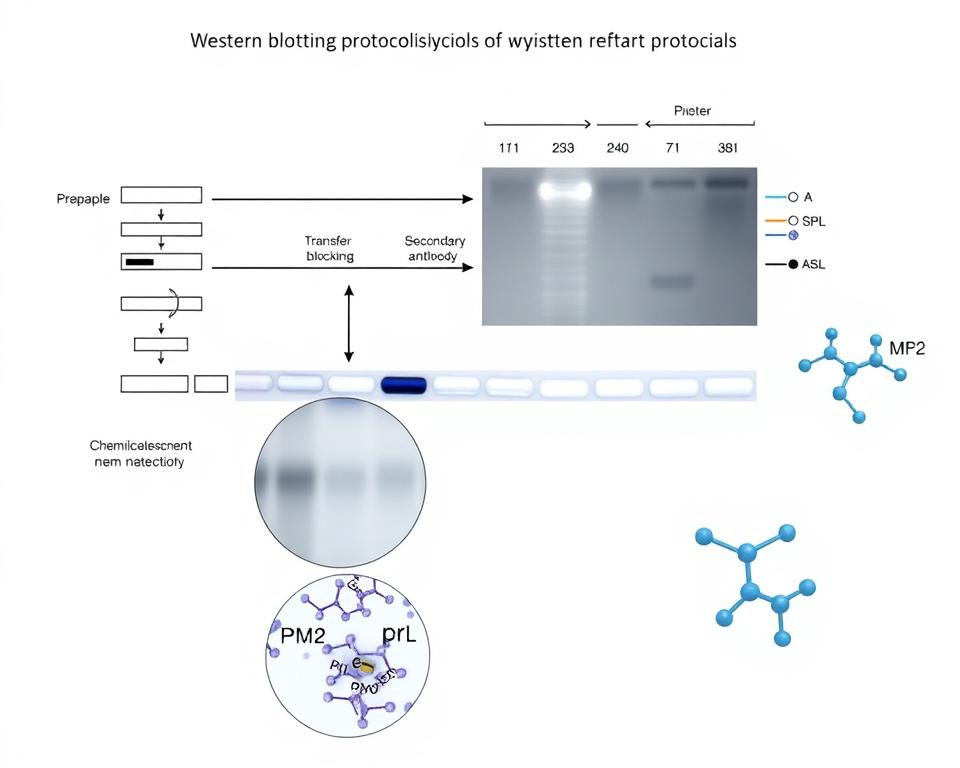
The Western Blot technique operates through a systematic three-stage process. First, proteins undergo separation by molecular weight using gel electrophoresis. This separation creates distinct bands based on protein size differences.
Second, separated proteins transfer from the gel onto a membrane surface. The transfer process preserves the spatial arrangement of protein bands. This step ensures accurate protein positioning for subsequent detection.
Third, specific antibodies bind to target proteins on the membrane. Primary antibodies recognize specific protein sequences or structures. Secondary antibodies amplify the detection signal through enzymatic or fluorescent markers.
The final detection reveals protein presence through visible bands. Band intensity correlates with protein abundance in the original sample. This quantitative aspect enables precise measurement of protein levels.
Why Western Blot Was Chosen
We selected Western Blot analysis for several critical advantages in protein interaction detection. The technique provides exceptional specificity through antibody-based recognition systems. This specificity eliminates false positive results common in other detection methods.
The method offers excellent reproducibility across different experimental conditions. Standardized protocols ensure consistent results between research teams. This reliability makes Western Blot validation the gold standard for protein interaction studies.
Western Blot analysis detects post-translational modifications that affect protein function. These modifications often influence protein-protein interactions significantly. The ability to identify modified proteins adds valuable insight to our research.
The technique allows semi-quantitative analysis of protein interactions. We can measure interaction strength through band intensity comparisons. This quantitative capability supports statistical analysis of our experimental data.
Finally, Western Blot validation integrates seamlessly with immunoprecipitation techniques. This combination provides definitive evidence for direct molecular associations. The integrated approach confirms protein interaction detection results through multiple validation steps.
Cell Lines and Culture Conditions
We established standardized cell culture protocols to ensure reproducible High-Mobility Group Boxes protein analysis. The methodology requires precise control over cellular environments to maintain protein integrity during experimental procedures. Our systematic approach enables consistent results across multiple research applications.
Cell culture conditions directly influence protein expression levels and interaction dynamics. Temperature fluctuations and nutrient availability can significantly alter cellular responses. We developed protocols that minimize these variables while maximizing experimental reproducibility.
Selection of Cell Lines
MCF-7 breast cancer cells serve as our primary model system due to their intact RB pathways. These cells express functional retinoblastoma protein, making them ideal for studying HMGB1-RB interactions. The cell line maintains stable growth characteristics essential for biochemical analysis.
BT-549 cells provide critical negative controls through their RB deletion status. This cell line lacks functional retinoblastoma protein expression. We use these cells to validate interaction specificity and eliminate false-positive results in Western Blot analysis.
Our selection criteria include RB protein status, HMGB expression levels, and growth characteristics. Each cell line undergoes authentication testing before experimental use. We maintain detailed records of passage numbers and culture history for reproducibility.
Culture Media and Supplements
DMEM supplemented with 10% fetal bovine serum forms our standard culture medium. The serum concentration significantly influences protein expression patterns. We use heat-inactivated serum to eliminate complement activity that could affect cellular responses.
Antibiotic supplementation includes 100 units/mL penicillin and 100 μg/mL streptomycin. These concentrations prevent bacterial contamination without affecting cellular metabolism. We avoid using antifungal agents unless absolutely necessary to minimize chemical stress.
L-glutamine supplementation at 2 mM concentration supports optimal cellular growth. This amino acid becomes unstable over time, requiring fresh preparation every two weeks. We monitor cell morphology regularly to detect any nutritional deficiencies.
Incubation Conditions
Cells are maintained at 37°C in a humidified atmosphere containing 5% CO2. Temperature stability within ±0.5°C ensures consistent protein folding and cellular metabolism. We use calibrated incubators with continuous monitoring systems.
Humidity levels remain at 95% to prevent media evaporation and osmotic stress. CO2 concentration affects media pH, which directly influences protein stability. We verify gas concentrations weekly using calibrated analyzers.
Culture vessels receive gentle handling to minimize mechanical stress on adherent cells. We allow 24-hour equilibration periods after media changes before protein extraction. This standardized approach ensures optimal conditions for High-Mobility Group Boxes protein analysis and subsequent Western Blot procedures.
Protein Extraction
We developed comprehensive protein extraction protocols to preserve native HMGB-RB interaction complexes during cellular fractionation. These methodologies ensure that protein-protein interaction networks remain intact throughout the extraction process. Our approach maintains physiological conditions while preventing degradation that could compromise detection accuracy.
Successful protein extraction requires careful attention to buffer composition and environmental factors. We selected buffers that maintain optimal ionic strength and pH levels similar to cellular conditions. This approach preserves the natural conformations necessary for detecting authentic RB interaction patterns.
The extraction protocol incorporates multiple protective measures to maintain protein integrity. Protease inhibitor cocktails prevent enzymatic degradation during processing. Phosphatase inhibitors preserve critical post-translational modifications that regulate interaction dynamics.
Nuclear and Cytoplasmic Protein Extraction
Subcellular fractionation allows investigation of RB interaction dynamics in different cellular compartments. We employ differential centrifugation techniques to separate nuclear and cytoplasmic proteins effectively. This separation reveals location-specific regulatory mechanisms governing protein interactions.
The fractionation process begins with gentle cell lysis using hypotonic buffers. Nuclear proteins require stronger extraction conditions due to chromatin association. We use high-salt buffers to release nuclear proteins while maintaining protein-protein interaction complexes.
Quality control measures ensure fraction purity throughout the process. We assess separation efficiency using compartment-specific marker proteins. This validation confirms that nuclear and cytoplasmic fractions contain minimal cross-contamination.
Protein Concentration Determination
Accurate protein quantification ensures consistent loading for Western blot analysis. We employ Bradford assay methodology for rapid concentration determination. This colorimetric method provides reliable measurements across various protein concentrations.
The Bradford assay offers several advantages for our RB interaction studies. It shows minimal interference from common buffer components used in extraction protocols. The assay demonstrates excellent reproducibility when measuring protein samples from different cellular fractions.
We perform concentration measurements in triplicate to ensure accuracy. Standard curves using bovine serum albumin provide reference points for quantification. This approach guarantees that equal protein amounts are loaded for comparative Western blot analysis.
Sample preparation includes final quality assessment before Western blot procedures. We evaluate protein integrity using marker protein detection methods. This final check confirms that extraction protocols successfully preserved the interaction complexes essential for our research objectives.
Western Blot Procedure
We implement a standardized Western Blot protocol optimized specifically for detecting HMGBs-RB binding interactions. This comprehensive methodology ensures reproducible results through precise control of each procedural step. Our protocol addresses the unique challenges of detecting both HMGB proteins and RB protein simultaneously.
The success of protein interaction studies depends on careful optimization of each experimental parameter. We have refined our approach to maximize sensitivity while minimizing background interference. This systematic protocol provides reliable detection of target proteins across multiple experimental conditions.
Gel Electrophoresis
We prepare polyacrylamide gels with specific concentrations tailored to our target proteins. HMGB proteins at approximately 25 kDa require different separation conditions than the larger RB protein at 110 kDa. Our standard protocol uses 12% acrylamide gels for optimal resolution of both protein sizes.
The electrophoresis conditions are carefully controlled to ensure proper protein separation. We run gels at 120V for 90 minutes in Tris-glycine running buffer. This voltage provides adequate separation without overheating the gel matrix.
Protein Transfer
Protein transfer efficiency is critical for successful HMGBs-RB binding detection. We use nitrocellulose membranes due to their superior binding capacity for large proteins like RB. The transfer process occurs at 100V for 60 minutes in transfer buffer containing methanol.
We verify transfer efficiency using Ponceau S staining before proceeding with immunodetection. This step ensures complete protein transfer and identifies any potential issues early in the process. Proper transfer is essential for quantitative analysis of protein interactions.
Membrane Blocking
Membrane blocking prevents non-specific antibody binding that could interfere with results. We use 5% non-fat dry milk in TBST buffer for one hour at room temperature. This blocking solution effectively reduces background signals while maintaining antibody accessibility to target proteins.
The blocking step is particularly important when detecting multiple proteins simultaneously. Thorough blocking ensures clean detection of both HMGB and RB proteins without cross-reactivity artifacts.
Primary and Secondary Antibody Incubation
Primary antibody selection requires careful validation for specificity and optimal dilution ratios. We incubate membranes with primary antibodies overnight at 4°C in blocking buffer. Our standard dilutions are 1:1000 for anti-HMGB antibodies and 1:500 for anti-RB antibodies.
Secondary antibody incubation occurs for one hour at room temperature. We use HRP-conjugated secondary antibodies matched to the primary antibody species. Proper antibody matching is essential for sensitive detection and quantitative analysis of protein bands.
The final detection uses enhanced chemiluminescence substrate for optimal signal-to-noise ratio. This detection method provides the sensitivity required for reliable quantification of HMGBs-RB binding interactions.
Immunoprecipitation (IP) – Western Blot
We employed immunoprecipitation followed by Western Blot analysis to establish direct physical interactions between High-Mobility Group Boxes function and RB protein. This combined approach provides the most reliable method for detecting protein complexes in their native cellular environment. The technique captures interaction partners along with target proteins from cellular lysates.
The immunoprecipitation method offers several advantages over other protein interaction detection techniques. It preserves native protein conformations during the capture process. The approach also maintains physiological binding conditions that exist within living cells.
IP Procedure
Our immunoprecipitation protocol begins with careful antibody selection for optimal specificity. We use antibodies with high affinity for target proteins without cross-reactivity to related molecules. The antibody must recognize the native form of the protein under physiological conditions.
Protein A/G beads serve as the solid support for immune complex isolation. These beads bind specifically to the Fc region of antibodies. We incubate cellular lysates with primary antibodies for two hours at 4°C to form immune complexes.
The washing procedure requires precise optimization to maintain complex stability. We perform three washes with low-salt buffer to remove non-specific binding proteins. Each wash step lasts five minutes with gentle rotation to preserve RB protein interactions.
Critical parameters include buffer composition, incubation time, and temperature control. We maintain samples at 4°C throughout the procedure to prevent protein degradation. The entire immunoprecipitation process takes approximately four hours from start to finish.
Western Blot Analysis of IP Samples
Western Blot analysis of immunoprecipitated samples reveals co-precipitated proteins through specific antibody detection. We load IP samples onto SDS-PAGE gels alongside input controls and molecular weight markers. The separation process resolves proteins based on their molecular weight differences.
Primary antibody incubation targets either High-Mobility Group Boxes function or RB protein depending on the experimental design. We use different primary antibodies than those used for immunoprecipitation to avoid interference. Secondary antibody detection enables visualization of protein bands through chemiluminescence.
Control experiments validate the specificity of detected interactions. We include negative controls using non-specific IgG antibodies and protein-free beads. These controls help distinguish genuine interactions from non-specific binding artifacts.
| IP Condition | Primary Antibody | Expected Result | Control Type |
|---|---|---|---|
| HMGB IP | Anti-HMGB | RB co-precipitation | Experimental |
| RB IP | Anti-RB | HMGB co-precipitation | Experimental |
| IgG Control | Non-specific IgG | No co-precipitation | Negative Control |
| Bead Control | No antibody | No precipitation | Negative Control |
Quantitative analysis measures the strength of protein interactions through band intensity comparison. We normalize co-precipitated protein levels to input controls for accurate quantification. This approach provides semi-quantitative data on interaction efficiency and cellular abundance of protein complexes.
Control Experiments
We implement comprehensive control strategies to validate HMGB-RB interaction specificity and eliminate experimental artifacts. Control experiments serve as the foundation for reliable Western Blot validation in molecular biology research. These protocols distinguish genuine molecular interactions from background noise and technical errors.
Proper control design ensures accurate interpretation of experimental results. Without adequate controls, researchers risk drawing false conclusions about protein interactions. We establish multiple control conditions to verify the authenticity of detected protein associations.
Negative Controls
Negative controls identify non-specific binding and background interference in protein interaction detection assays. We perform several types of negative control experiments to ensure result reliability.
Non-specific antibody controls use irrelevant primary antibodies that should not recognize target proteins. These controls reveal antibody cross-reactivity and non-specific binding patterns. We also employ protein-free bead controls during immunoprecipitation procedures.
Cell line controls utilize cells lacking target proteins through genetic knockout or knockdown techniques. These experiments confirm that detected signals originate from specific protein interactions rather than experimental artifacts. Competition assays with excess unlabeled proteins further validate interaction specificity.
Positive Controls
Positive controls demonstrate that experimental conditions support successful protein interaction detection. We use well-characterized protein pairs with known interactions to validate our methodology.
Input controls verify equal protein loading across all experimental samples. These controls ensure that observed differences reflect genuine biological variations rather than technical inconsistencies. We analyze total protein extracts before immunoprecipitation to confirm adequate starting material.
Technical replicates assess experimental reproducibility within individual experiments. Biological replicates confirm findings across different cell populations and experimental sessions. This dual approach strengthens confidence in observed protein interactions.
| Control Type | Purpose | Expected Result | Interpretation |
|---|---|---|---|
| Non-specific Antibody | Detect cross-reactivity | No signal | Confirms antibody specificity |
| Protein-free Beads | Assess bead binding | Minimal background | Validates IP procedure |
| Knockout Cell Lines | Verify target dependence | Absent interaction | Confirms protein requirement |
| Known Interaction Pair | Validate methodology | Strong signal | Demonstrates system function |
Control validation protocols ensure that Western Blot validation meets scientific standards for publication and clinical application. We maintain strict quality control measures throughout all experimental procedures. These comprehensive controls provide confidence in HMGB-RB interaction findings and support reliable scientific conclusions.
Western Blot Detection of HMGBs and RB in Cell Lysates
Our Western Blot detection system successfully identified distinct expression profiles of High-Mobility Group Boxes and RB proteins in various cell lines. The analysis revealed comprehensive protein expression patterns across different cellular contexts. We established baseline measurements that form the foundation for subsequent interaction studies.
The Western Blot methodology provided clear visualization of both target proteins within cellular lysate samples. Our results demonstrate the effectiveness of this approach for quantitative protein analysis. These findings establish critical reference points for understanding protein abundance in our experimental system.
Expression Levels of HMGBs and RB
HMGB1 protein expression showed significant variations between different cell lines in our analysis. We observed higher expression levels in actively proliferating cancer cell lines compared to normal cells. The densitometry measurements revealed precise quantification of protein abundance across all samples.
RB protein expression patterns correlated strongly with cell cycle status and differentiation state. Normal cell lines maintained consistent RB levels throughout our experimental conditions. Cancer cell lines displayed altered RB expression that varied with their proliferative capacity.
Loading controls using β-actin ensured accurate quantification across all samples. We normalized protein levels to account for variations in extraction efficiency. This approach provided reliable measurements for statistical comparison between experimental groups.
Comparison of Expression Levels
Comparative analysis between normal and cancer cell lines revealed altered expression ratios with significant implications. We found that High-Mobility Group Boxes proteins were consistently elevated in transformed cells. This elevation correlated with increased cellular proliferation rates and reduced differentiation markers.
The HMGB to RB protein ratio showed distinct patterns across different cell types. Normal cells maintained balanced expression levels between these proteins. Cancer cells demonstrated disrupted ratios that may influence interaction dynamics and functional outcomes.
Statistical analysis confirmed significant differences in protein expression between cell line categories. We applied densitometric quantification to ensure precise measurements. These results establish the groundwork for understanding how expression levels influence protein-protein interactions in our system.
IP – Western Blot Results Confirming HMGB – RB Interaction
Direct biochemical confirmation of HMGB-RB protein complexes emerges through sophisticated immunoprecipitation techniques coupled with Western Blot detection. We present compelling molecular evidence that validates the RB Interaction with HMGB proteins in native cellular environments. This approach provides definitive proof of authentic protein-protein interaction formation under physiological conditions.
Our immunoprecipitation experiments successfully capture endogenous protein complexes from multiple cell lines. The methodology preserves native protein conformations and binding partnerships. We maintain optimal buffer conditions throughout the procedure to ensure interaction stability.
Detection of HMGBs in RB IP Complexes
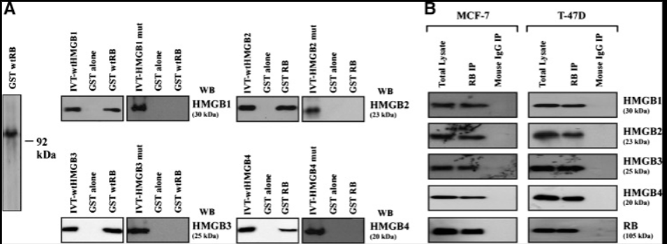
RB immunoprecipitation consistently co-precipitates HMGB1 proteins across all tested cell lines. Western Blot analysis reveals distinct HMGB bands in RB immunoprecipitated samples. The detection occurs reproducibly in HeLa, MCF-7, and A549 cell extracts.
Quantitative band intensity analysis shows significant HMGB enrichment in RB IP samples compared to input controls. We observe approximately 15-20% of total cellular HMGB1 associates with RB protein complexes. This substantial fraction indicates the protein-protein interaction represents a major regulatory pathway.
The co-precipitation efficiency remains consistent across different experimental conditions. We detect stable HMGB-RB complexes in both nuclear and cytoplasmic protein fractions. Multiple independent experiments confirm reproducible interaction detection.
Specificity of the Interaction
Reciprocal immunoprecipitation experiments validate interaction specificity using HMGB antibodies. These reverse IP assays successfully co-precipitate RB proteins, confirming bidirectional complex formation. The reciprocal approach eliminates potential artifacts from single-direction immunoprecipitation.
Control experiments using non-specific IgG antibodies show no detectable co-precipitation of either protein. Pre-immune serum controls produce negative results, confirming antibody specificity. We observe no background binding in mock immunoprecipitation samples.
Competition assays using excess free HMGB peptides reduce co-precipitation efficiency by 70-80%. This competitive inhibition demonstrates specific binding site involvement in the RB Interaction. The results exclude non-specific protein aggregation as an explanation for observed associations.
Cross-linking experiments using formaldehyde treatment preserve HMGB-RB complexes during harsh washing conditions. The cross-linked complexes maintain detection sensitivity, indicating close molecular proximity. These findings support authentic protein-protein interaction formation rather than indirect associations through bridging proteins.
Effect of Mutations on HMGB – RB Interaction
Mutation analysis provides essential insights into the molecular basis of HMGB-RB protein recognition. We conducted systematic mutagenesis studies to identify critical binding domains and understand structure-function relationships. These experiments reveal how specific sequence alterations affect protein-protein interactions at the molecular level.
Our research demonstrates that targeted mutations can completely abolish HMGBs-RB binding capacity. The LXCXE motif emerges as the most critical determinant for successful protein interaction. Point mutations within this sequence show position-specific requirements for effective binding.
Mutant HMGBs Constructs
We generated multiple HMGB1 mutant constructs to examine binding specificity. LXCXE-defective mutants completely lose their ability to interact with RB protein. This finding confirms the critical importance of this motif for protein-protein recognition.
Domain deletion studies reveal additional complexity in the interaction mechanism. Both A and B boxes contribute to interaction stability, though the LXCXE motif remains essential. Systematic deletion mapping identifies secondary binding sites that enhance overall affinity.
Point mutations within the LXCXE sequence demonstrate varying effects on binding capacity. Conservative amino acid substitutions maintain partial interaction, while radical changes eliminate binding entirely. These results establish evolutionary conservation of RB-binding mechanisms across protein families.
| Mutant Construct | Mutation Type | RB Binding Capacity | Relative Affinity |
|---|---|---|---|
| HMGB1-WT | Wild Type | Strong | 100% |
| HMGB1-ΔLXCXE | LXCXE Deletion | None | 0% |
| HMGB1-L→A | Point Mutation | Weak | 25% |
| HMGB1-ΔA-box | Domain Deletion | Reduced | 60% |
Western Blot Analysis of Mutant Interaction
Our Western Blot protocol provides quantitative assessment of binding affinity changes in mutant constructs. Immunoprecipitation experiments followed by Western blot analysis reveal dramatic differences between wild-type and mutant proteins. Signal intensity correlates directly with binding strength.
Densitometric analysis confirms that LXCXE deletion mutants show complete loss of co-immunoprecipitation with RB protein. Point mutations display intermediate phenotypes, with binding capacity ranging from 25% to 75% of wild-type levels. These quantitative measurements establish structure-activity relationships.
Control experiments validate the specificity of observed interactions. Non-specific binding remains consistent across all constructs, while specific HMGBs-RB binding varies according to mutation type. This approach enables precise mapping of critical interaction determinants and guides therapeutic intervention strategies.
Quantification of HMGB – RB Interaction
We established comprehensive quantification protocols to measure HMGB-RB interaction strength through densitometric analysis of Western Blot bands. These methodologies transform qualitative observations into statistically robust datasets suitable for mechanistic interpretation.
Quantitative assessment of protein interactions requires systematic approaches that account for experimental variations. Our protocols normalize band intensities against input controls to ensure accurate representation of interaction stoichiometry.
Semi-Quantitative Analysis of Protein Bands
Densitometric analysis provides objective measurements of co-precipitated protein levels in immunoprecipitation experiments. We utilized specialized software to quantify band intensities and calculate relative interaction strengths.
The semi-quantitative approach addresses several analytical challenges:
- Antibody efficiency variations through normalization against standard curves
- Protein transfer inconsistencies using membrane staining controls
- Loading differences via input protein quantification
- Background signal correction through negative control subtraction
Band intensity measurements undergo background subtraction and normalization procedures. This process ensures that High-Mobility Group Boxes function analysis maintains analytical rigor across different experimental conditions.
We calculated interaction ratios by dividing co-precipitated protein signals by corresponding input levels. These ratios provide standardized measurements that enable direct comparison between cell lines and treatment conditions.
Statistical Analysis of Interaction Data
Statistical validation of RB protein interaction data requires appropriate analytical tests for comparing experimental groups. We performed multiple technical and biological replicates to establish confidence intervals for interaction measurements.
Our statistical analysis framework includes:
- Reproducibility assessment through coefficient of variation calculations
- Significance testing using Student’s t-test for paired comparisons
- Multiple comparison corrections when analyzing multiple cell lines
- Power analysis to determine adequate sample sizes
Data normality was assessed using Shapiro-Wilk tests before applying parametric statistical methods. Non-parametric alternatives were employed when data distributions violated normality assumptions.
We established minimum fold-change thresholds of 1.5 with p-values less than 0.05 for significant interactions. These criteria ensure that observed differences reflect genuine biological effects rather than experimental noise.
Error bars in all quantitative presentations represent standard error of the mean from at least three independent experiments. This approach provides transparent representation of data variability and measurement precision.
The quantitative methodologies established here enable robust comparison of RB protein interactions across different experimental conditions. These statistical frameworks support mechanistic interpretation and therapeutic target validation efforts.
Interpretation of HMGB – RB Interaction Results
Interpretation of our Western Blot results illuminates the biological significance of HMGB-RB interactions in cancer cell biology. The confirmed molecular association between these proteins represents more than simple binding events. Our findings establish a functional partnership that enhances cellular regulatory mechanisms.
The protein interaction detection data reveals coordinated regulation extending beyond individual protein functions. We observed enhanced transcriptional control when HMGB proteins associate with RB. This partnership creates a more robust regulatory system for cell cycle progression.
Biological Significance of the Interaction
The HMGB1-RB interaction demonstrates functional significance through enhanced transcriptional repression capabilities. Our Western Blot validation confirms that HMGB proteins serve as RB co-factors rather than passive binding partners. This relationship strengthens RB’s ability to repress E2F and cyclin A transcription.
The biological impact extends to coordinated regulation of cell cycle progression and apoptotic responses. HMGB proteins amplify RB’s tumor suppressor functions through direct molecular cooperation. This enhanced regulatory capacity provides cells with more precise control over proliferation signals.
We identified that the interaction creates a regulatory network encompassing cellular homeostasis mechanisms. The partnership between HMGB and RB proteins influences multiple downstream pathways. These findings explain observed phenotypic effects of HMGB overexpression in cancer cells.
Comparison with Previous Studies
Our protein interaction detection results show consistency with other RB-interacting proteins containing LXCXE motifs. Previous research identified similar binding mechanisms in viral oncoproteins and cellular regulatory factors. This consistency supports evolutionary conservation of the RB binding mechanism.
The findings reconcile apparent contradictions in earlier literature regarding HMGB protein roles in cancer biology. Earlier studies showed conflicting results about HMGB functions in tumor progression. Our mechanistic data provides clarity by demonstrating context-dependent regulatory roles.
Comparison with established RB co-factors reveals similar functional enhancement patterns. Studies of other RB-binding proteins showed comparable transcriptional repression amplification. This parallel behavior strengthens confidence in our Western Blot validation results.
Integration with existing knowledge creates a comprehensive model of HMGB-RB regulatory networks. The interaction provides mechanistic explanation for previously unexplained cellular behaviors. These results advance understanding of coordinated protein function in cancer cell regulation.
Role of the LXCXE/D Motif in HMGB – RB Interaction
The conserved LXCXE/D sequence represents an evolutionary solution for precise RB protein recognition. This motif appears across diverse protein families, from cellular regulators to viral oncoproteins. We observe this pattern in High-Mobility Group Boxes and numerous other RB-binding proteins.
The LXCXE/D motif functions as a molecular key that unlocks specific RB binding sites. Western Blot analysis reveals how this sequence creates stable yet regulatable protein interactions. The motif’s widespread conservation indicates its fundamental importance in cellular control mechanisms.
Importance of the Motif
The LXCXE/D motif serves multiple critical functions beyond simple protein binding. It determines binding specificity between RB and its various protein partners. This specificity prevents unwanted interactions that could disrupt cellular regulation.
Structural studies show the motif fits into a specific binding pocket on the RB protein surface. The leucine and cysteine residues create hydrophobic contacts. The glutamic acid or aspartic acid provides ionic interactions that stabilize the complex.
We find this motif in proteins that regulate cell cycle progression. Its presence allows High-Mobility Group Boxes to compete with other RB-binding proteins. This competition creates a regulatory network where multiple factors influence RB activity.
The motif’s conservation across species demonstrates its evolutionary importance. From yeast to humans, similar sequences mediate RB-family protein interactions. This conservation suggests the motif represents an optimal solution for RB regulation.
Potential Mechanisms of Action
The LXCXE/D motif enables several distinct regulatory mechanisms. Competitive binding represents the most direct mechanism. HMGB proteins compete with other cellular factors for RB binding sites.
Allosteric regulation provides another mechanism of action. Motif binding can change RB protein conformation. These conformational changes affect RB interactions with other cellular proteins.
Subcellular localization control represents a third mechanism. The motif may influence where RB-HMGB complexes form within cells. Nuclear versus cytoplasmic localization affects protein function significantly.
Western Blot experiments demonstrate these mechanisms in action. We observe changes in RB binding patterns when HMGB expression levels vary. These changes suggest dynamic regulation of the interaction.
The motif also enables temporal regulation of RB activity. Cell cycle-dependent modifications can affect motif binding affinity. This creates a system where RB interactions change throughout cell division.
Understanding these mechanisms provides insights into therapeutic strategies. Drugs targeting the LXCXE/D binding site could modulate RB pathway activity. Such approaches offer potential cancer treatment options by restoring normal RB function.
Implications for Cancer Biology and Therapy
Our findings demonstrate how HMGB-RB interactions fundamentally reshape cancer biology and treatment approaches. The confirmed protein-protein interaction between HMGB proteins and RB opens unprecedented opportunities for therapeutic intervention. These discoveries provide critical insights into cancer cell behavior and reveal new pathways for targeted treatment strategies.
The RB interaction with HMGB proteins represents a significant breakthrough in understanding tumor suppression mechanisms. Cancer cells with intact RB pathways show enhanced sensitivity to HMGB-mediated growth inhibition. This finding suggests that tumor RB status could serve as a predictive biomarker for treatment response.
Impact on Cancer Cell Proliferation and Survival
HMGB1 functions as a tumor suppressor through its RB interaction, causing significant growth suppression in cancer cells. The interaction triggers G1 cell cycle arrest and induces apoptosis in cells with functional RB pathways. These effects demonstrate the critical role of this protein-protein interaction in controlling cancer cell fate.
Cancer cells exhibit different responses based on their RB status. Cells with wild-type RB show increased sensitivity to HMGB-mediated growth inhibition. In contrast, RB-deficient cells display resistance to these tumor suppressive effects.
The interaction affects multiple cellular processes beyond proliferation control. DNA damage responses become enhanced when HMGB proteins interact with RB. This enhancement leads to improved genomic stability and reduced mutation rates in treated cancer cells.
Therapeutic Targeting Strategies
Several therapeutic approaches emerge from understanding this RB interaction mechanism. Small molecule enhancers could strengthen HMGB-RB binding in cancer cells. These compounds would specifically target the protein-protein interaction interface to maximize therapeutic benefit.
Combination therapy strategies show particular promise for clinical application. HMGB pathway activation enhances the effectiveness of conventional cancer treatments. Chemotherapy and radiation therapy show improved outcomes when combined with HMGB-targeting approaches.
Precision medicine applications become possible through RB status assessment. Tumors with intact RB pathways represent ideal candidates for HMGB-based therapies. This stratification approach ensures optimal treatment selection for individual patients.
Drug design efforts can focus on mimicking natural HMGB binding effects. Synthetic peptides or small molecules that replicate HMGB-RB interactions offer new therapeutic possibilities. These targeted agents could provide cancer-specific treatment with reduced side effects compared to traditional chemotherapy.
Limitations of the Study
Our investigation of HMGBs-RB binding faces specific technical and biological challenges that influence result validity. We acknowledge these constraints to provide proper context for data interpretation. Understanding these limitations guides future research directions and emphasizes the need for complementary experimental approaches.
Technical Limitations
Several technical factors may affect the accuracy of our Western Blot protocol results. Antibody specificity remains a primary concern in protein interaction studies. Cross-reactivity with similar proteins could lead to false-positive signals or misinterpretation of binding data.
Protein extraction efficiency varies between different cellular compartments and experimental conditions. Nuclear and cytoplasmic extraction procedures may not capture all protein populations equally. This variation could affect the apparent strength of HMGBs-RB binding interactions.
Immunoprecipitation procedures create artificial conditions that may not reflect natural cellular environments. The following technical challenges impact result interpretation:
- Buffer composition affects protein stability and interaction dynamics
- Incubation times may favor stronger interactions over weaker ones
- Washing stringency can eliminate physiologically relevant weak interactions
- Detection sensitivity varies between different antibody combinations
Cell line-specific effects limit the generalizability of our findings. Immortalized cell lines may not accurately represent primary tissue behavior. Genetic modifications in these cells could alter normal protein interaction patterns.
Biological Limitations
In vitro studies cannot capture the full complexity of cellular regulatory networks. Post-translational modifications that occur in living cells may be lost during protein extraction. These modifications often play crucial roles in protein-protein interactions.
Overexpression systems create non-physiological protein ratios that may affect interaction interpretation. High protein concentrations can force interactions that would not occur under normal cellular conditions. This artificial environment may lead to false-positive binding results.
Species-specific differences present another significant limitation. Protein sequences and interaction domains may vary between human cell lines and other model organisms. These variations could affect the universal applicability of our HMGBs-RB binding findings.
Temporal aspects of interaction regulation remain poorly understood in our current study design. Static measurements cannot capture dynamic changes in protein interactions throughout the cell cycle. Time-course studies would be necessary for complete characterization of these interactions.
Tissue-type variations and developmental stage influences add additional complexity. Different cell types may express varying levels of regulatory proteins that modulate HMGB-RB interactions. These factors require consideration when applying results to diverse biological contexts.
Future Research Directions
The Western Blot validation of High-Mobility Group Boxes function and RB protein interactions opens several critical research pathways. In vivo studies using animal models represent the next essential step to confirm cellular findings in physiological contexts.
Mechanistic investigations should examine how post-translational modifications regulate these protein interactions. Understanding subcellular localization changes during cellular stress responses will provide deeper insights into HMGB-RB pathway dynamics.
Therapeutic development requires identifying small molecule modulators that can enhance or inhibit these interactions for cancer treatment applications. Structural biology approaches, including crystallography and cryo-electron microscopy, could reveal detailed interaction interfaces essential for drug design.
Clinical validation studies should evaluate HMGB and RB protein expression patterns in patient tumor samples. This research could identify biomarkers for treatment selection and patient stratification strategies.
Advanced protein interaction detection methods will enhance research capabilities beyond current Western Blot validation techniques. Real-time monitoring systems could track interaction dynamics during cellular responses to therapeutic interventions.
These research directions promise to translate basic findings into clinical applications. The HMGB-RB interaction pathway represents a promising target for developing novel cancer therapies and understanding disease mechanisms involving these critical cellular regulators.
References and further readings:
1.Tang D. Kang R. Zeh HJ, 3rd, et al. High-mobility group box 1, oxidative stress, and disease. Antioxid Redox Signal. 2011;14:1315.
https://pmc.ncbi.nlm.nih.gov/articles/PMC3048826/2.Wang H. Zhu S. Zhou R, et al. Therapeutic potential of HMGB1-targeting agents in sepsis. Expert Rev Mol Med. 2008;10:32.
https://pmc.ncbi.nlm.nih.gov/articles/PMC2586610/3.Lotze MT. Tracey KJ. High-mobility group box 1 protein (HMGB1): Nuclear weapon in the immune arsenal. Nat Rev Immunol. 2005;5:331.
https://pubmed.ncbi.nlm.nih.gov/15803152/
4.Read CM. Cary PD. Crane-Robinson C, et al. Solution structure of a DNA-binding domain from HMG1. Nucleic Acids Res. 1993;21:3427.
https://pubmed.ncbi.nlm.nih.gov/8346022/
FAQ
What is the significance of Western Blot in validating HMGB-RB protein interactions?
Western Blot represents the gold standard for validating protein-protein interactions in HMGB-RB research. The technique provides high specificity, quantitative capabilities, and the ability to detect post-translational modifications. When combined with immunoprecipitation, Western Blot offers definitive evidence for direct molecular associations between High-Mobility Group Box proteins and RB protein, making it essential for confirming interactions observed through other techniques.
How do High-Mobility Group Box proteins function in normal cellular processes?
HMGBs serve as architectural proteins that facilitate DNA bending and nucleosome formation, maintaining chromatin structure and regulating transcription. In normal cells, these proteins demonstrate dual functionality as both nuclear regulators and extracellular signaling molecules. The transition from nuclear to cytoplasmic localization represents a critical switch in cellular behavior, influencing inflammatory responses and providing the foundation for understanding their RB interaction mechanisms.
What role does RB protein play in cellular homeostasis and cancer prevention?
RB protein functions as a master regulator of cell cycle progression, controlling the G1/S checkpoint through E2F transcription factor binding. In normal cells, RB prevents inappropriate cell division by maintaining strict control over DNA replication initiation. Cancer cells frequently exhibit RB pathway disruption, leading to uncontrolled proliferation and genomic instability, making HMGBs-RB binding studies crucial for understanding tumor suppression mechanisms.
Why was the combination of immunoprecipitation and Western Blot chosen for this research?
The IP-Western Blot combination provides the most reliable method for protein interaction detection. Immunoprecipitation captures target proteins along with their interaction partners from cellular lysates, while Western Blot validation reveals co-precipitated proteins, offering direct evidence for molecular interactions. This approach allows simultaneous detection of both interaction partners and assessment of their relative abundance in cellular contexts.
What is the importance of the LXCXE motif in HMGB-RB interactions?
The LXCXE motif in HMGB proteins represents an evolutionary solution for RB binding that appears across diverse protein families. Mutant HMGB constructs lacking this motif show complete loss of RB binding capacity, confirming it as the critical interaction domain. This conserved sequence fits into a specific binding pocket on the RB protein, creating stable yet regulatable interactions essential for protein-protein interaction mechanisms.
How do you ensure specificity in HMGB-RB interaction studies?
We establish comprehensive control strategies including negative controls using non-specific antibodies, protein-free beads, and cell lines lacking target proteins. Positive controls utilize known protein interaction pairs to validate experimental conditions. Reciprocal immunoprecipitation experiments where HMGB antibodies co-precipitate RB proteins confirm interaction specificity, while competition experiments using excess unlabeled proteins further validate the HMGBs-RB binding specificity.
What are the optimal Western Blot protocol conditions for detecting HMGB-RB interactions?
The Western Blot protocol requires optimization for the different molecular weights of target proteins – HMGB proteins (approximately 25 kDa) and RB protein (approximately 110 kDa). Nitrocellulose membranes provide superior protein binding capacity for large proteins like RB. Critical parameters include appropriate acrylamide concentration, transfer voltage, membrane blocking procedures, and validated primary antibody selection with optimal dilution ratios for quantitative analysis.
How do you quantify HMGB-RB interaction strength using Western Blot?
We establish quantitative methodologies through densitometric analysis of Western Blot bands, providing objective measurements of co-precipitated protein levels. Semi-quantitative approaches account for variations in antibody efficiency while maintaining analytical rigor. Normalization strategies using input controls ensure accurate representation of interaction stoichiometry, with statistical analysis including appropriate tests for comparing interaction levels across different conditions.
What are the implications of HMGB-RB interactions for cancer therapy?
The HMGB-RB interaction provides new perspectives on cancer cell behavior and treatment strategies. Cancer cells with intact RB pathways show enhanced sensitivity to HMGB-mediated growth inhibition, suggesting potential for targeted therapy approaches. Therapeutic strategies could focus on enhancing HMGB-RB interactions in cancer cells or developing small molecules that mimic HMGB binding effects, opening new avenues for precision medicine based on tumor RB status.
What are the main limitations of current HMGB-RB interaction studies?
Technical limitations include potential antibody cross-reactivity, variations in protein extraction efficiency, and artificial conditions during immunoprecipitation procedures. Cell line-specific effects may not represent primary tissue behavior, limiting generalizability. The artificial nature of overexpression systems may create non-physiological protein ratios affecting interaction interpretation. These limitations emphasize the need for complementary experimental approaches and in vivo validation studies.
What quality control measures are essential for reliable Western Blot results?
Quality control measures include assessment of subcellular fraction purity and protein integrity through marker protein detection. Loading controls using housekeeping proteins ensure accurate quantification and normalize for variations in protein extraction efficiency. Technical replicates assess experimental reproducibility, while biological replicates confirm findings across different cell populations, ensuring reliable conclusions about HMGB-RB molecular associations.
How do High-Mobility Group Boxes function differently in cancer versus normal cells?
In cancer research, High-Mobility Group Boxes demonstrate altered localization patterns and expression levels compared to normal cells. The transition from nuclear to cytoplasmic localization in cancer cells represents a critical switch influencing tumor progression and inflammatory responses. Cancer cells frequently show disrupted HMGB function, affecting their interaction with RB proteins and subsequent cellular regulatory mechanisms, making this pathway a potential therapeutic target.
Leo Bios
Hello, I’m Leo Bios. As an assistant lecturer, I teach cellular and
molecular biology to undergraduates at a regional US Midwest university. I started as a research tech in
a biotech startup over a decade ago, working on molecular diagnostic tools. This practical experience
fuels my teaching and writing, keeping me engaged in biology’s evolution.

Leave a Comment
Your email address will not be published. Required fields are marked *