


When protein detection fails in a western blot, it can be frustrating. We will explore the common causes of this issue and provide practical solutions to optimize your results.
Western blotting is a fundamental analytical technique used to identify specific proteins within complex mixtures. The process involves separation by size, transfer to a membrane, and detection using antibodies. Issues with any of these steps can lead to a lack of signal.
To troubleshoot, we need to systematically examine each step of the protocol. Common issues include problems with antibody binding, gel or membrane issues, and errors in sample preparation or detection methods.
Key Takeaways
- Understand the basic principles of western blotting to effectively troubleshoot signal issues.
- Common causes of signal failure include issues with protein transfer, antibody-related problems, and detection method failures.
- Systematically examining each step of the western blot protocol is crucial for identifying the cause of signal failure.
- Optimizing the western blot process involves verifying each step, from sample preparation to detection.
- Practical solutions are available to fix common issues and improve western blot results.
Understanding Western Blot Technique
Understanding the principles of western blotting is essential for researchers to accurately detect and quantify proteins within complex biological samples. Western blotting is a multi-step analytical technique that involves the separation, transfer, and detection of proteins using specific antibodies.
Basic Principles of Western Blotting
The basic principles of western blotting involve three fundamental steps: separation of proteins by size using gel electrophoresis, transfer of these proteins to a solid membrane support, and detection of specific proteins using antibodies. In the first step, proteins are denatured and separated based on their molecular weight through sodium dodecyl sulfate-polyacrylamide gel electrophoresis (SDS-PAGE).
Key Components for Successful Western Blot
Key components for successful western blot include high-quality protein samples, appropriate gel selection, efficient transfer systems, specific primary antibodies, sensitive detection methods, and optimized blocking and washing protocols. The quality and specificity of antibodies used are particularly crucial, as they directly determine the sensitivity and specificity of protein detection in the final analysis.
Protein Transfer Issues: The Most Common Culprit
The efficiency of protein transfer from the gel to the membrane is crucial for successful Western blotting. Protein transfer issues are a significant challenge, as they directly impact the detection of target proteins.
Incomplete Transfer to Membrane
Incomplete transfer to the membrane can occur due to several factors, including improper assembly of the transfer “sandwich,” air bubbles between the gel and membrane, incorrect buffer composition, or inadequate transfer time. Large molecular weight proteins (>100 kDa) often require extended transfer times or specialized transfer conditions.
The composition of the transfer buffer is critical; insufficient methanol can reduce binding of proteins to nitrocellulose membranes, while too much SDS can cause proteins to pass through the membrane without binding. Proper assembly of the transfer apparatus is essential, ensuring that the gel and membrane are in complete contact and correctly oriented relative to the electrodes.
How to Verify Successful Protein Transfer
To verify successful protein transfer, researchers can use reversible protein stains such as Ponceau S to visualize all proteins on the membrane before proceeding with antibody incubation. Alternatively, pre-stained molecular weight markers provide a visual confirmation that at least some proteins have transferred from the gel to the membrane.
For a more thorough verification, researchers can stain the post-transfer gel with Coomassie blue to confirm that proteins have indeed migrated out of the gel. Optimizing transfer conditions based on protein size—using wet transfer for larger proteins and semi-dry transfer for smaller proteins—can significantly improve transfer efficiency and subsequent signal detection.
For more detailed information on optimizing Western blotting techniques, you can refer to our comprehensive guide on Western Blotting Technology.
Antibody-Related Problems Causing No Signal
Antibody-related issues are a significant cause of signal failure in Western blotting experiments. The choice of primary antibody and secondary antibody plays a crucial role in the detection of the target protein. When these antibodies are not optimized for the specific application, it can lead to a lack of signal or non-specific binding.
Primary Antibody Concentration and Specificity Issues
The concentration and specificity of the primary antibody are critical factors that influence the outcome of a Western blot. If the primary antibody is too dilute, it may not bind sufficiently to the target protein, resulting in a weak or absent signal. Conversely, if it is too concentrated, it can cause non-specific binding and high background noise. Moreover, the specificity of the primary antibody to the protein of interest is paramount. An antibody that recognizes a different epitope than the one present in the denatured protein will fail to generate a signal.
To avoid these issues, researchers should verify that the primary antibody has been validated for Western blotting applications with the specific species and cell type being studied. Not all antibodies are suitable for Western blotting; some work well for other techniques like immunoprecipitation or immunohistochemistry but fail in Western blots because they recognize conformational epitopes lost during denaturation.
Secondary Antibody Selection and Dilution Mistakes
The secondary antibody must be precisely matched to the host species of the primary antibody. For instance, if the primary antibody is derived from a mouse, the secondary antibody should be an anti-mouse immunoglobulin. Common mistakes include using incorrect species reactivity, improper dilution, or choosing a detection system that is incompatible with the available imaging equipment.
The age and storage conditions of antibodies can also significantly impact their performance. Antibodies that have undergone multiple freeze-thaw cycles or been stored improperly may lose activity and fail to generate a signal. When troubleshooting antibody issues, including positive controls can help verify that the antibody detection system is functioning properly.
Sample Preparation Errors Leading to Signal Loss
Sample preparation is a critical step in western blot analysis, and errors in this process can lead to signal loss. We will explore common mistakes made during sample preparation and how they impact the success of western blot detection.
Protein Degradation During Extraction
Protein degradation during extraction is a significant issue that can result in signal loss. If samples are not kept cold throughout the extraction process or if protease inhibitors are omitted from lysis buffers, proteins can degrade rapidly. This degradation can lead to fragmented proteins that may no longer be recognized by antibodies, thus reducing the signal. To mitigate this, it’s essential to maintain samples on ice and include protease inhibitors in the lysis buffer.
Different sample types require optimized cell lysis conditions. For instance, tissue samples often necessitate more aggressive mechanical disruption methods like homogenization or sonication, whereas cultured cells may be lysed with milder conditions. The composition of lysis buffers should be tailored to the cellular location of the protein of interest. For example, membrane proteins require detergent-based buffers, while cytosolic proteins can be extracted with milder conditions.
Improper Sample Loading and Normalization
Improper sample loading can result from inaccurate protein quantification, leading to inconsistent amounts of protein across gel lanes. This inconsistency can potentially result in insufficient target protein for detection, thus causing signal loss. Normalization errors occur when unequal amounts of protein are loaded across samples, making it impossible to make valid comparisons between experimental conditions.
To ensure accurate sample loading, it’s crucial to measure protein concentration using a spectrophotometer. The appropriate volume of the sample should then be diluted into a loading buffer containing glycerol and a tracking dye, such as bromophenol blue. The sample is then heated to denature the higher-order structure while retaining disulfide bridges.
| Sample Preparation Step | Common Error | Impact on Western Blot |
|---|---|---|
| Protein Extraction | Omission of protease inhibitors | Protein degradation, signal loss |
| Sample Loading | Inaccurate protein quantification | Inconsistent protein amounts, signal variability |
| Sample Denaturation | Insufficient or excessive heating | Protein aggregation or incomplete denaturation |
By understanding and addressing these common sample preparation errors, you can significantly improve the reliability and consistency of your western blot results.
Western Blot Detection Method Failures
The success of western blotting depends heavily on the detection method used. Detection method failures represent a significant source of signal problems in western blotting, particularly when using enzyme-linked detection systems such as horseradish peroxidase (HRP) or alkaline phosphatase (AP).
Chemiluminescence Detection Problems
Chemiluminescence detection, while highly sensitive, is prone to several technical issues that can result in no signal. Expired or improperly stored detection reagents, enzyme inactivation, and improper mixing of substrate components are common problems. The quality and freshness of chemiluminescent substrates are critical, as these reagents typically have limited shelf lives and may lose activity if stored improperly or used beyond their expiration dates.
Enzyme conjugates on secondary antibodies can become inactivated through improper storage, repeated freeze-thaw cycles, or contamination with sodium azide, a common preservative that inhibits HRP activity. To avoid these issues, it’s essential to store detection reagents according to the manufacturer’s instructions and to use them within their specified shelf life.
Substrate Depletion and Exposure Time Issues
Substrate depletion can occur rapidly in areas of high protein concentration, leading to localized signal loss even when the detection system is functioning properly in other regions of the membrane. Optimizing exposure time is also crucial for chemiluminescent detection. Too short an exposure may fail to capture weak signals, while excessive exposure can lead to film saturation that masks true signal differences.
Digital imaging systems, while increasingly popular, have their own set of potential failure points, including improper calibration, insufficient dynamic range, or software settings that filter out weak signals. The timing between substrate application and image acquisition is critical for chemiluminescent detection, as the signal intensity typically peaks within minutes and then gradually declines.
To troubleshoot detection failures, it’s advisable to test the detection system independently using dot blots with dilutions of secondary antibody to verify that the detection reagents are functioning properly. By understanding these potential pitfalls and taking steps to mitigate them, researchers can optimize their western blotting protocols to achieve reliable and consistent results.
Blocking Buffer and Washing Protocol Issues
Optimizing the blocking buffer and washing protocol is essential for achieving reliable Western blot results. The choice of blocking agent and the conditions under which the membrane is washed significantly impact the signal-to-noise ratio and the specificity of antibody binding.
Inappropriate Blocking Agent Selection
The selection of an appropriate blocking agent is critical and must be compatible with both the membrane type (nitrocellulose or PVDF) and the detection system. Non-fat dry milk is commonly used due to its cost-effectiveness and availability. However, it contains biotin and phosphoproteins that can interfere with certain detection systems, particularly those using streptavidin-based detection or anti-phosphoprotein antibodies. In such cases, BSA (Bovine Serum Albumin) is preferred as it does not contain these interfering components.
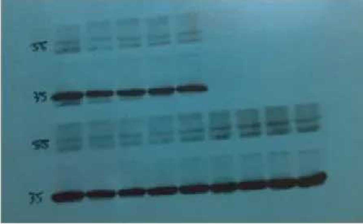
weak-signal bands
The concentration of the blocking agent is also crucial. Too dilute solutions may not adequately prevent non-specific binding, while overly concentrated solutions can mask epitopes and prevent the primary antibody from binding to the target protein. The blocking time must also be optimized to balance between sufficient blocking and signal preservation.
Insufficient or Excessive Washing Steps
The washing steps are vital for removing unbound antibodies and minimizing background noise. However, excessive washing can strip specifically bound antibodies from the membrane, especially if the detergent concentration is too high or the washing time is too long. The type of membrane used influences the washing requirements; PVDF membranes typically require more stringent conditions due to their higher protein-binding capacity compared to nitrocellulose membranes.
To optimize the washing protocol, the concentration of detergent (such as Tween-20) in the washing buffer must be carefully adjusted. Insufficient washing can leave excess unbound antibodies, resulting in high background, while excessive washing can reduce the signal by stripping away specifically bound antibodies.
Step-by-Step Troubleshooting Protocol for No Signal
Identifying the cause of no signal in western blots demands a thorough and systematic troubleshooting process. When you encounter a western blot with no visible signal, it’s essential to methodically examine each step of the protocol to pinpoint the issue.
Systematic Approach to Identify the Cause
To troubleshoot effectively, start by verifying protein transfer to the membrane using reversible stains like Ponceau S. This step helps determine if the issue lies with the transfer process. Next, examine each step of the western blot protocol sequentially, including sample preparation, gel electrophoresis, transfer efficiency, blocking conditions, antibody incubations, and finally, detection methods.
A systematic approach involves checking the gel for proper loading and ensuring that the antibody concentrations are appropriate. It’s also crucial to verify that the transfer buffer and other reagents are not contaminated.
| Troubleshooting Step | Action | Expected Outcome |
|---|---|---|
| Verify Protein Transfer | Use Ponceau S staining | Visible protein bands on the membrane |
| Check Antibody Functionality | Perform dot blot with purified protein | Signal detection indicates functional antibody |
| Assess Detection System | Apply HRP-conjugated antibody directly to the membrane | Signal production confirms detection reagents are working |
Control Experiments to Pinpoint the Problem
Control experiments are vital for identifying the exact source of the problem. Include positive controls (samples known to contain the protein of interest) and loading controls (housekeeping proteins like β-actin or GAPDH) in every western blot. Test the functionality of the primary antibody using dot blots with purified protein or lysates known to express high levels of the target protein.
Verify secondary antibody activity by performing a dot blot with only the secondary antibody on a membrane spotted with the appropriate immunoglobulin. Assess the detection system independently by applying a small amount of HRP-conjugated antibody directly to the membrane and developing with substrate.
By following this systematic troubleshooting protocol and utilizing control experiments, you can effectively identify and resolve the issue causing the lack of signal in your western blot.
Preventative Measures for Consistent Western Blot Results
Implementing quality control checkpoints is vital for successful western blotting. To achieve reliable results, it’s essential to establish standardized protocols for each step of the western blotting process, including sample preparation, gel electrophoresis, transfer conditions, blocking, antibody incubations, and detection methods.
Quality Control Checkpoints Throughout the Protocol
To ensure the integrity of your western blot results, incorporate quality control checkpoints at critical stages. Verify protein concentration after extraction, confirm proper gel polymerization before loading samples, and check transfer efficiency before proceeding with antibody incubations. These checks help identify potential issues early on, allowing for timely corrections.
- Verify protein concentration after extraction to ensure accurate sample loading.
- Confirm proper gel polymerization before loading samples to prevent gel-related issues.
- Check transfer efficiency to ensure that proteins have been successfully transferred to the membrane.
| Quality Control Checkpoint | Purpose | Action |
|---|---|---|
| Protein concentration verification | Ensure accurate sample loading | Use a spectrophotometer or Bradford assay |
| Gel polymerization check | Prevent gel-related issues | Visually inspect the gel for proper polymerization |
| Transfer efficiency check | Ensure successful protein transfer | Use Ponceau S staining or a similar method |
Best Practices for Reagent Storage and Handling
Proper storage and handling of reagents are crucial for maintaining their activity and ensuring consistent western blot results. Aliquot antibodies into single-use volumes to avoid repeated freeze-thaw cycles, and store them according to manufacturer recommendations, typically at -20°C or -80°C for long-term storage.
- Store antibodies at the recommended temperature to maintain their activity.
- Prepare fresh transfer and running buffers for each experiment to prevent buffer degradation.
- Maintain detailed records of reagent preparation dates, lot numbers, and expiration dates to track potential sources of variability.
By implementing these preventative measures and best practices, you can significantly improve the reliability and consistency of your western blot results.
Conclusion: Ensuring Reliable Western Blot Signals
In conclusion, mastering the western blot technique requires a deep understanding of its intricacies and potential pitfalls. Western blotting remains one of the most powerful and widely used techniques for protein analysis in molecular biology, but its success depends on careful optimization and troubleshooting of each step in the protocol.
The five most common causes of no signal in western blots—protein transfer issues, antibody-related problems, sample preparation errors, detection method failures, and blocking/washing protocol issues—can all be systematically addressed through the approaches outlined in this article. By understanding and addressing these common issues, researchers can significantly improve their western blot results.
To achieve reliable western blot signals, it’s essential to implement preventative measures and quality control checkpoints throughout the protocol. This includes careful sample preparation, optimal antibody selection, and precise detection methods. By doing so, you can significantly reduce the frequency of signal failures and improve the consistency of your western blot results.
Remember that western blotting is a semi-quantitative technique, and results should be interpreted with caution. With proper attention to detail and systematic optimization, western blotting can provide reliable, reproducible results that advance our understanding of protein expression and function in biological systems.
References and further readings:
1.Sule, R., Rivera, G., & Gomes, A. V. (2023). Western blotting (immunoblotting): history, theory, uses, protocol and problems. BioTechniques.
https://www.tandfonline.com/doi/full/10.2144/btn-2022-00342.Kurien, B. T., & Scofield, R. H. (2006). Western blotting. Methods, 38(4), 283–293.
https://www.sciencedirect.com/science/article/abs/pii/S10462023060000653.Eaton, S. L., Hurtado, M. L., & Oldknow, K. J., et al. (2014). A guide to modern quantitative fluorescent western blotting with troubleshooting strategies. Journal of Visualized Experiments, 93, e52099.
https://pmc.ncbi.nlm.nih.gov/articles/PMC4354265/
FAQ
What are the most common causes of no signal in a protein detection experiment?
The most common causes include issues with protein transfer, antibody specificity, sample preparation, and detection methods. We recommend checking the gel electrophoresis and transfer buffer conditions, as well as verifying the primary and secondary antibody concentrations and specificity.
How can I verify successful protein transfer to the membrane?
To verify successful protein transfer, you can use Ponceau S staining or Coomassie blue staining on the membrane after transfer. Alternatively, you can perform a reversible staining using a compatible dye. We also recommend checking the transfer apparatus and buffer composition to ensure optimal conditions.
What is the optimal concentration for primary and secondary antibodies?
The optimal concentration for primary and secondary antibodies varies depending on the specific antibodies and experimental conditions. We recommend starting with the manufacturer’s recommended dilution and optimizing as needed. Typically, primary antibody dilutions range from 1:500 to 1:1000, while secondary antibody dilutions range from 1:1000 to 1:5000.
How can I minimize protein degradation during sample preparation?
To minimize protein degradation, we recommend using protease inhibitors during sample extraction and handling samples on ice or at 4°C. Additionally, using a suitable lysis buffer and optimizing the sample loading and normalization protocols can help prevent protein degradation.
What are the best practices for storing and handling Western blot reagents?
To ensure optimal performance, we recommend storing antibodies, buffers, and other reagents according to the manufacturer’s instructions. Typically, antibodies should be stored at -20°C or 4°C, while buffers should be prepared fresh or stored at 4°C. Handling reagents in a sterile environment and avoiding contamination can also help maintain their quality.
How can I troubleshoot issues with chemiluminescence detection?
To troubleshoot chemiluminescence detection issues, we recommend checking the substrate and exposure time. Ensure that the substrate is not depleted and that the exposure time is optimized for signal detection. Additionally, verifying the secondary antibody and detection reagent compatibility can help resolve detection issues.
Leo Bios
Hello, I’m Leo Bios. As an assistant lecturer, I teach cellular and
molecular biology to undergraduates at a regional US Midwest university. I started as a research tech in
a biotech startup over a decade ago, working on molecular diagnostic tools. This practical experience
fuels my teaching and writing, keeping me engaged in biology’s evolution.

Leave a Comment
Your email address will not be published. Required fields are marked *