SKU: WM0171 Categories: ,

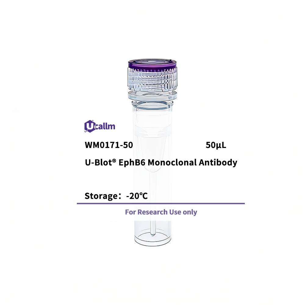
EphB6 Monoclonal Antibody

Price range: $268.00 through $328.00

SKU: WM0171-50
Enter the quantity you would like, or drag the panel to find the quantity and price that best for you
Bulk QuoteAuthorize OEM & ODM

Product details

Background:
This gene encodes a member of a family of transmembrane proteins that function as receptors for ephrin-B family proteins. Unlike other members of this family, the encoded protein does not contain a functional kinase domain. Activity of this protein can influence cell adhesion and migration. Expression of this gene is downregulated during tumor progression, suggesting that the protein may suppress tumor invasion and metastasis. Alternative splicing results in multiple transcript variants. [provided by RefSeq, Jul 2013],


Specifications

Size50μL/100μL
TargetEphB6
ReactivityHuman
ApplicationsWB, IHC, IF, ELISA
MW(Calculated)111kD
MW(Observed)
Host SpeciesMouse
Isotype
Conjugate/ModificationUnmodified
Recommended Dilution RatioWB 1:500-1:2000; IHC 1:200-1:1000; ELISA 1:10000; IF 1:50-200 / Note: For IHC, we suggest antigen retrieval with TE buffer pH 9.0 (Cat#WT1004)
FormulationLiquid in PBS containing 50% glycerol, 0.5% BSA and 0.02% sodium azide.
PurificationAffinity purification
Storage-15°C to -25°C/1 year(Do not lower than -25°C)
Concentration
ClonalityMonoclonal
Clone Number6H8
ImmunogenPurified recombinant fragment of EphB6 expressed in E. Coli.
SpecificityEphB6 Monoclonal Antibody detects endogenous levels of EphB6 protein.
Gene NameEPHB6
Protein NameEphrin type-B receptor 6
Other NameEPHB6; / Ephrin type-B receptor 6; / HEP; / Tyrosine-protein kinase-defective receptor EPH-6
SpeciesHuman
Gene ID2051
UniprotO15197,
Species.1Mouse
Gene ID.1
Uniprot.1O08644
Species.2
Gene ID.2
Uniprot.2
Cellular LocalizationMembrane; Single-pass type I membrane protein.; [Isoform 3]: Secreted .
Tissue ExpressionExpressed in brain. Expressed in non invasive breast carcinoma cell lines (at protein level). Strong expression in brain and pancreas, and weak expression in other tissues, such as heart, placenta, lung, liver, skeletal muscle and kidney. Expressed in breast non invasive tumors but not in metastatic lesions. Isoform 3 is expressed in cell lines of glioblastomas, anaplastic astrocytomas, gliosarcomas and astrocytomas. Isoform 3 is not detected in normal tissues.
Signaling_pathway
Research Areas>>Axon guidance
FunctionDomain:The protein kinase domain is predicted to be catalytically inactive. Its extracellular domain is capable of promoting cell adhesion and migration in response to low concentrations of ephrin-B2, but its cytoplasmic domain is essential for cell repulsion and inhibition of migration induced by high concentrations of ephrin-B2.,Function:Kinase-defective receptor for members of the ephrin-B family. Binds to ephrin-B1 and ephrin-B2. Modulates cell adhesion and migration by exerting both positive and negative effects upon stimulation with ephrin-B2. Inhibits JNK activation, T cell receptor-induced IL-2 secretion and CD25 expression upon stimulation with ephrin-B2.,PTM:Ligand-binding increases phosphorylation on tyrosine residues. Phosphorylation on tyrosine residues is mediated by transphosphorylation by the catalytically active EPHB1 in a ligand-independent manner. Tyrosine phosphorylation of the receptor may act as a switch on the functional transition from cell adhesion/attraction to de-adhesion/repulsion.,similarity:Belongs to the protein kinase superfamily. Tyr protein kinase family. Ephrin receptor subfamily.,similarity:Contains 1 protein kinase domain.,similarity:Contains 1 SAM (sterile alpha motif) domain.,similarity:Contains 2 fibronectin type-III domains.,subunit:Interacts with CBL and EPHB1. Interacts with FYN; this interaction takes place in a ligand-independent manner.,tissue specificity:Expressed in brain. Expressed in non invasive breast carcinoma cell lines (at protein level). Strong expression in brain and pancreas, and weak expression in other tissues, such as heart, placenta, lung, liver, skeletal muscle and kidney. Expressed in breast non invasive tumors but not in metastatic lesions. Isoform 3 is expressed in cell lines of glioblastomas, anaplastic astrocytomas, gliosarcomas and astrocytomas. Isoform 3 is not detected in normal tissues.,
Please login to post questions
Back to Top
Product has been added to your cart