Ki-67 Polyclonal Antibody
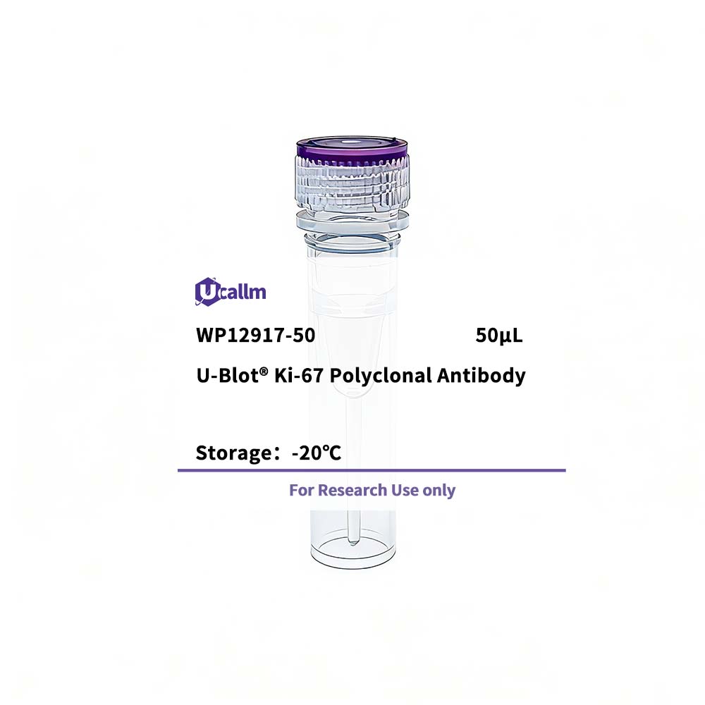
Ucallm_WP12917-50
Price range: $268.00 through $328.00
Product details
Background:
This gene encodes a nuclear protein that is associated with and may be necessary for cellular proliferation. Alternatively spliced transcript variants have been described. A related pseudogene exists on chromosome X. [provided by RefSeq, Mar 2009],
Specifications
| Size | 50μL/100μL |
| Target | Ki-67 |
| Reactivity | Human |
| Applications | IHC, IF, ELISA |
| MW(Calculated) | |
| MW(Observed) | 360kD |
| Host Species | Rabbit |
| Isotype | IgG |
| Conjugate/Modification | Unmodified |
| Recommended Dilution Ratio | IHC 1:100-1:300; IF 1:200-1:1000; ELISA 1:20000; Not yet tested in other applications. / Note: For IHC, we suggest antigen retrieval with TE buffer pH 9.0 (Cat#WT1004) |
| Formulation | Liquid in PBS containing 50% glycerol, 0.5% BSA and 0.02% sodium azide. |
| Purification | The antibody was affinity-purified from rabbit antiserum by affinity-chromatography using epitope-specific immunogen. |
| Storage | -15°C to -25°C/1 year(Do not lower than -25°C) |
| Concentration | 1 mg/ml |
| Clonality | Polyclonal |
| Clone Number | |
| Immunogen | The antiserum was produced against synthesized peptide derived from human Ki67. AA range:3207-3256 |
| Specificity | Ki-67 Polyclonal Antibody detects endogenous levels of Ki-67 protein. |
| Gene Name | MKI67 |
| Protein Name | Ki 67 |
| Other Name | MKI67; / Antigen KI-67 |
| Species | Human |
| Gene ID | 4288 |
| Uniprot | P46013 |
| Species.1 | |
| Gene ID.1 | |
| Uniprot.1 | |
| Species.2 | |
| Gene ID.2 | |
| Uniprot.2 | |
| Cellular Localization | Chromosome . Nucleus . Nucleus, nucleolus . Associates with the surface of the mitotic chromosome, the perichromosomal layer, and covers a substantial fraction of the mitotic chromosome surface (PubMed:27362226). Associates with satellite DNA in G1 phase (PubMed:9510506). Binds tightly to chromatin in interphase, chromatin-binding decreases in mitosis when it associates with the surface of the condensed chromosomes (PubMed:15896774, PubMed:22002106). Predominantly localized in the G1 phase in the perinucleolar region, in the later phases it is also detected throughout the nuclear interior, being predominantly localized in the nuclear matrix (PubMed:22002106). . |
| Tissue Expression | Epithelium, |
| Signaling_pathway | |
| Research Areas | |
| Function | developmental stage:Expression of this antigen occurs preferentially during late G1, S, G2 and M phases of the cell cycle, while in cells in G0 phase the antigen cannot be detected.,Function:Thought to be required for maintaining cell proliferation.,online information:Ki-67 entry,similarity:Contains 1 FHA domain.,subcellular location:Predominantly localized in the G1 phase in the perinucleolar region, in the later phases it is also detected throughout the nuclear interior, being predominantly localized in the nuclear matrix. In mitosis, it is present on all chromosomes.,subunit:Interacts with KIF15. Binds through the FHA domain to MKI67IP., |
Please login to post questions
Related products