Ueasy® Bis-Tris precast gels

Ueasy® precast gels are polyacrylamide electrophoresis gels, mainly used for protein separation.It has two core specification classifications:By gel concentration: gradient gels (4-12% and 4-20%) and fixed-concentration gels (8%, 10% and 12%);By well number: 12-well and 15-well.Sample Loading Specification12-well gel: Maximum loading volume 50 uL, recommended loading volume within 25 uL.15-well gel: Maximum loading volume 30 uL, recommended loading volume within 15 uL.

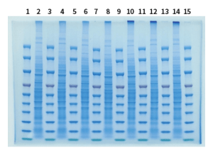
Uniformity Test

Gel:W3M25(4-20%、15 wells)

Voltage:120 V

Time:80 min

Lane1、3、5、7、9、11、13、15:Prestained Protein Ladder(W2100)4 uL

Lane2、4、6、8、10、12、14:BSA 4 uL(20 ug)


Resolution Test

Gel:W3M25(4-20%、15 wells)

Voltage:120 V

Time:80 min

Lane1、15:restained Protein Ladder(W2100)4 uL

Lane2-14:BSA 20 uL(20 ug)

Same Sample at Different Voltages

Purpose:

Electrophoresis tests of the same sample at different voltages were performed to comprehensively validate the core performance of the precast gels. This verifies electrophoresis compatibility, minimizes interference from gel efficiency variations, defines the full voltage tolerance range, ensures experimental repeatability, and eliminates issues such as uneven bands or smearing caused by voltage fluctuations, resulting in more controllable experimental outcomes.

Stable bands at low voltage confirm uniform pore size of the precast gels, making them suitable for long-duration low-speed electrophoresis with excellent separation repeatability.

Normal bands at high voltage demonstrate outstanding mechanical strength, enabling resistance to heat and current stress generated during high-speed electrophoresis, with stable and reliable performance.
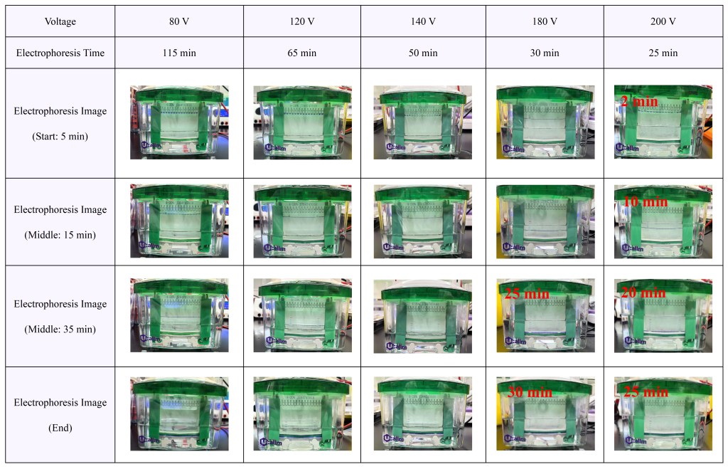
Ueasy® Bis-Tris precast gels(4-20% 15wells)

Electrophoresis Images of the Same Sample at Different Voltages
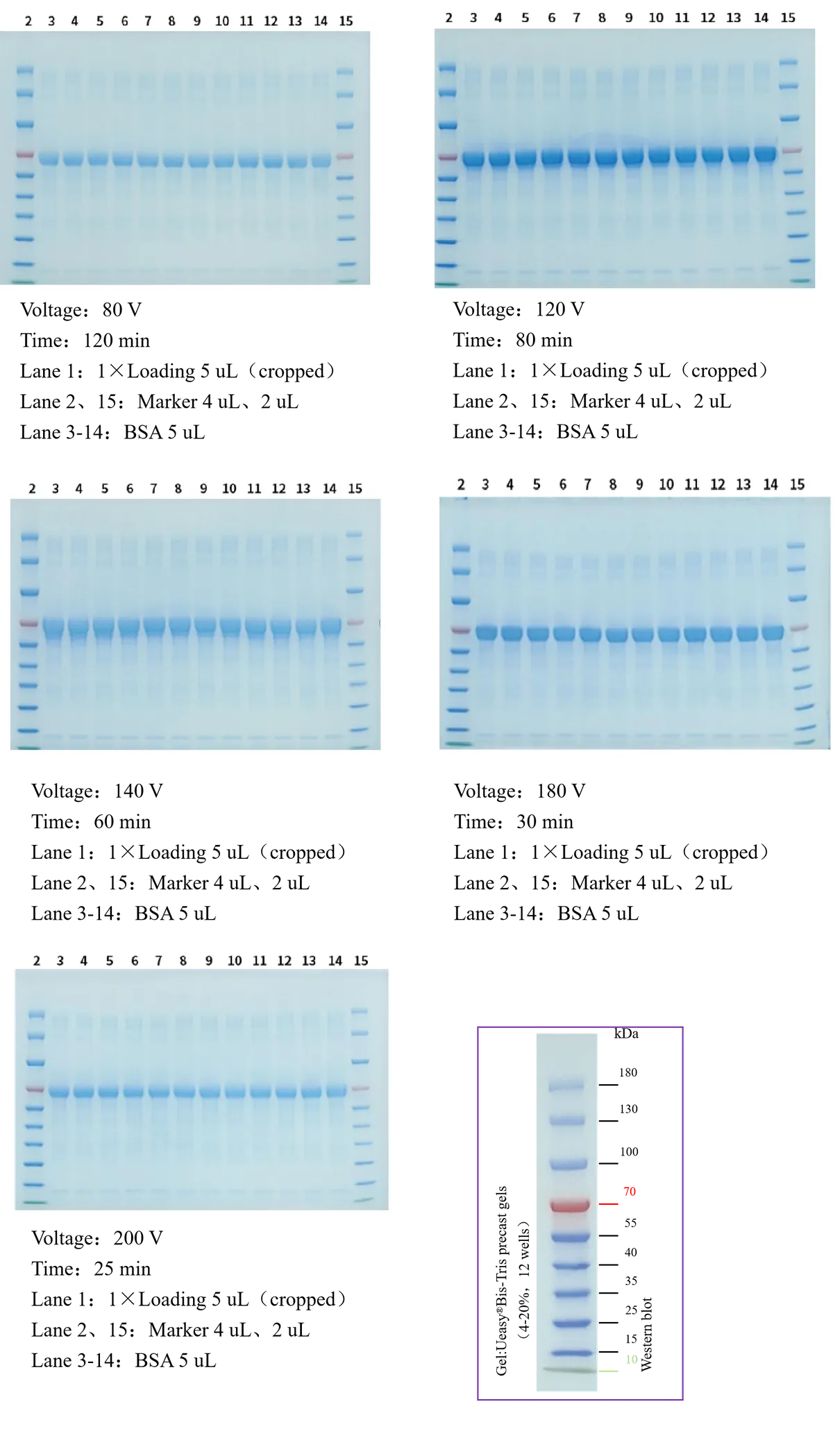
Result Analysis:

At room temperature (25 ℃), electrophoresis time decreases with increasing voltage for Ueasy® Bis-Tris precast gels. Although inner chamber temperature rises gradually during electrophoresis, it remains within the applicable range of the gel. Under all tested voltages, target protein bands are clear and Marker migration rates are consistent with no obvious deviation.

Purpose:

This experiment tests precast gels using the same sample with different loading volumes and electrophoresis voltages. The two main objectives are:

  • To determine the optimal loading volume to avoid band distortion.
  • To identify the optimal voltage that ensures band quality while shortening electrophoresis time and improving efficiency.

Insufficient loading: Low protein content leads to faint, weak bands or undetectable signals.Excessive loading: Protein overload causes band broadening, smearing, increased non‑specific bands, and interference with accurate Marker estimation.
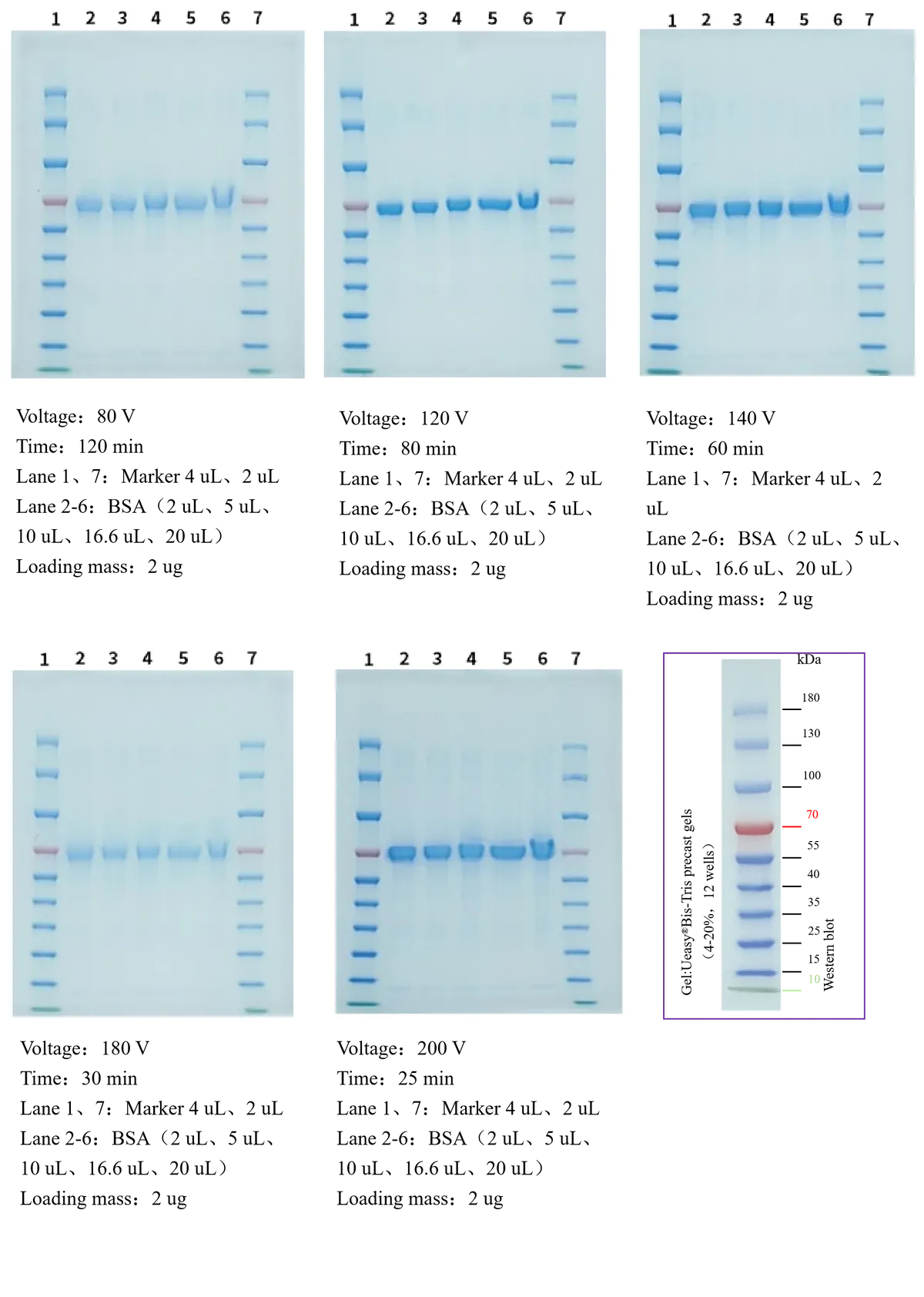
Result Analysis:

At room temperature (25 ℃), electrophoresis time decreases and inner chamber temperature rises moderately (within safe limits) with increasing voltage for Ueasy® Bis-Tris precast gels. Protein bands remain normal at 2 uL, 5 uL, 10 uL, and 16.6 uL loading volumes. The 20 uL loading shows smearing, blurred edges, and band broadening at all voltages, indicating overload and well retention leading to distorted bands.

Purpose:

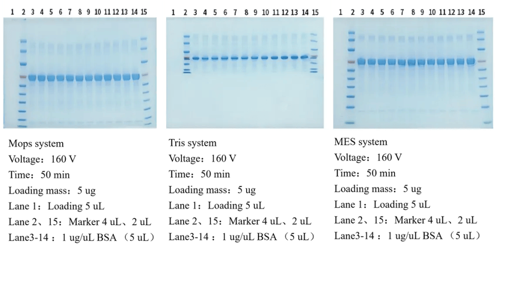
This experiment uses samples of identical concentration and volume to evaluate precast gels in different electrophoresis systems. The goal is to optimize protein separation and select the best system for the target protein molecular weight.MOPS, Tris-Glycine, and MES buffers differ in ionic strength and pH, leading to distinct molecular weight separation ranges. Consistent loading allows direct comparison to achieve efficient separation, avoid band overlap, diffusion, and abnormal migration.

MOPS system: pH 7.2-7.8, strong buffering capacity, uniform migration, reduces smearing of medium-to-high molecular weight proteins.

Tris-Glycine system: Classic, pH-8.3, stable, cost-effective, ideal for initial screening of unknown molecular weights.

MES system: pH 5.5-6.7, fast separation, sharp compact bands, minimizes diffusion and overlap of low molecular weight proteins; improves band shape in acidic conditions.

Result Analysis:

At 25℃, Ueasy® Bis-Tris precast gels were tested in three buffer systems. MOPS and MES showed similar separation speed and longer migration distance with comparable overall performance. Tris-Glycine achieved only~50% migration progress, with significantly lower separation efficiency.

Purpose:

To evaluate the effect of high-salt environments on protein separation in precast gels. High-salt samples (cell lysates, body fluids containing high NaCl, KCl, etc.) introduce excess ions that compete with SDS for protein binding, disrupt uniform charge, alter buffer ionic strength, and cause gel pore shrinkage or swelling. This leads to aberrant migration, smearing, diffusion, and Marker misalignment.

Result Analysis:

At 25℃, high-salt samples in Ueasy® Bis-Tris precast gels show significantly slower migration and wider wells compared to the control. High salt alters ionic strength, causing abnormal gel pore changes and disturbed migration. Smearing, blurred edges, and high background are also observed, indicating competition between salt ions and SDS, disrupting protein charge uniformity and uniform migration.

Purpose:

The core objectives are to directly verify protein separation efficiency, rationality of loading volume, and gel performance.

(1) Verify the separation resolution of the electrophoresis system for proteins at different concentrationsBSA is an ideal standard protein due to its uniform molecular weight and high purity. By loading gradient concentrations of BSA for electrophoresis combined with Coomassie brilliant blue staining, the separation performance of the electrophoresis system can be visually and quantitatively evaluated. The specific criteria are as follows:

Clear and distinguishable bands for low-concentration BSA → indicates that the electrophoresis system meets the sensitivity requirement for trace protein separation and can satisfy the detection of low-abundance proteins.

Sharp, non-smearing, and non-diffusing bands for high-concentration BSA → proves good pore adaptability of the precast gels, which avoids separation failure caused by protein overload and meets the experimental tolerance range.

Uniform spacing and no overlap among bands of different BSA concentrations → demonstrates scientific setting of core electrophoresis conditions (voltage, running time, gel concentration), a good linear relationship between protein migration rate and concentration, and excellent separation stability.

(2) Calibrate the loading volume to avoid abnormal band risks in subsequent WB experimentsGel staining is a direct method to visually judge the appropriateness of loading volume. The loading parameters can be reversely calibrated according to BSA band morphology to avoid deviations in subsequent WB caused by improper loading. The specific calibration logic is as follows:

Overly wide bands or “smiling bands” for high-concentration BSA → indicates overloading. The loading volume should be reduced in subsequent experiments; otherwise, it will lead to uneven membrane transfer and increased non-specific antibody binding.

Faint or invisible bands for low-concentration BSA → indicates insufficient loading. The loading volume should be increased to avoid weak target signals and false-negative results in WB.

(3) Evaluate gel quality and stability of electrophoresis operationThe stained gel results of BSA at different concentrations can be directly used as a validation basis for gel performance and operational standardization. The specific evaluation criteria are as follows:

Consistent position and intensity of duplicate lanes for the same BSA concentration → proves stable gel batches, standardized casting, no bubbles or uneven pores, and qualified core performance.

Abnormalities such as skewed bands, blurred edges, or uneven intensity → indicates problems during electrophoresis, possibly caused by unstable voltage, poor sealing of gel plates, or expired buffer. Experimental conditions should be adjusted promptly to ensure the reliability of subsequent experiments.
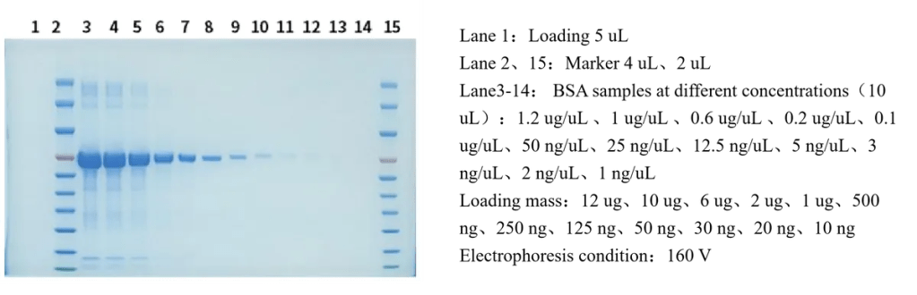
Result Analysis:

At room temperature (25 °C), twelve gradient protein concentrations (1.2 μg/μL, 1 μg/μL, 0.6 μg/μL, 0.2 μg/μL, 0.1 μg/μL, 50 ng/μL, 25 ng/μL, 12.5 ng/μL, 5 ng/μL, 3 ng/μL, 2 ng/μL, 1 ng/μL) were tested using Ueasy® Bis-Tris precast gels. The results showed that the first three high-concentration gradients (1.2–0.6 μg/μL) were excessively high and not recommended for subsequent detection. No obvious bands were observed when the protein concentration was below 2 ng/μL, so the protein concentration should be no lower than this threshold in follow-up tests.

1.Electrophoresis-Related Problems

Q1:After opening a new precast gel, the electrophoresis bands appear skewed or uneven at the edges. What are the causes?

  • The main causes and solutions are as follows:
  1. Inconsistent sample loading volumes: Large differences in loading volume across lanes lead to uneven liquid tension during electrophoresis.
  2. Unlevel or leaking electrophoresis tank: The tank is not level, the buffer leaks, or the buffer levels in the inner and outer chambers are inconsistent. Before use, level the tank and ensure the buffer levels are equal and there is no leakage.

Q2:How to resolve smearing, diffusion, or low resolution after electrophoresis?

  • This issue is mostly related to sample handling, electrophoresis conditions, or gel storage:
  1. Insufficient sample purity: High salt concentration, contaminating proteins, or nucleic acids in the sample cause band smearing. Desalt the sample or treat it with nuclease to degrade nucleic acids.
  2. Improper voltage/current: Excessively high voltage causes gel heating and protein diffusion; excessively low voltage leads to incomplete separation.
  3. Expired or improperly stored precast gel: Degraded acrylamide polymerization affects separation performance. Check the expiration date. Store unopened gels at 2–8 °C in the dark, and avoid prolonged exposure to room temperature.

Q3: What to do if the wells of the precast gel collapse or leak, preventing normal sample loading?

  • Causes and solutions:
  1. Incorrect comb removal: Pulling the comb at an angle or with excessive force deforms the wells. Remove the comb slowly in a vertical direction. If wells are slightly deformed, gently reshape the edges with a pipette tip.
  2. Gel drying: Prolonged exposure after opening causes the well gel to shrink. Assemble the electrophoresis unit immediately after opening. If not using immediately, add a small amount of electrophoresis buffer to the wells to keep them moist.

Q4:After electrophoresis with the precast gel, the target protein fails to transfer efficiently to the membrane. What are the causes?

  • The main causes are related to the matching between gel concentration and transfer conditions:
    1. Mismatch between gel concentration and protein molecular weight: Our 4-20% precast gels are generally suitable for the separation of proteins across common molecular weights. For very low-molecular-weight proteins, use dedicated precast gels, as otherwise the small pore size will hinder transfer. For very high-molecular-weight proteins, low-concentration gels (6%–8%) may bind proteins too tightly for efficient transfer. Select the appropriate gel concentration according to the molecular weight of the target protein.
    2. Insufficient transfer time or current: If the transfer time is too short or the current is too low, the protein will not be fully transferred.
    3. Inactive transfer buffer: Excessively high methanol concentration (>20%) will overfix proteins in the gel; excessively low concentration will cause the gel to swell easily. Use fresh transfer buffer and maintain the methanol concentration at 10%–20%.

Q5:After electrophoresis with the precast gel, there are excessive non-specific bands and high background. How to optimize?

  • Non-specific bands and high background are related to the gel, sample, and antibody. Optimization directions are as follows:
    1. Insufficient washing of the precast gel: Residual ions or stabilizers in the gel cause background staining. After electrophoresis, rinse the gel with transfer buffer to remove residual impurities.
    2. Excessively high primary antibody concentration or prolonged incubation time: Reduce the primary antibody concentration (recommended dilution: 1:5000–1:10000) and shorten the incubation time.
    3. Inadequate blocking: First, ensure the blocking solution completely covers the membrane. Second, appropriately extend the blocking time. Most importantly, select an appropriate blocking reagent.

Q6:What is the shelf life of the precast gels?

  • Standard precast gels (e.g., SDS-PAGE gels) stored at 2-8 °C in the dark typically

have a shelf life of 6-12 months (refer to the manufacturer’s instructions for specific details).

Q7:Can the same batch of electrophoresis buffer be used with precast gels from different brands?

  • Mixing is not recommended. Formulations of precast gels (e.g., cross-linking degree, stabilizer type) vary between brands, leading to different requirements for buffer ionic strength and pH. Mixing buffers may reduce electrophoresis efficiency and cause abnormal band patterns. It is recommended to use the buffer formulation specified by the precast gel manufacturer or the matching electrophoresis buffer from the same brand.

 Q8:Can electrophoresis continue after the bromophenol blue dye reaches the bottom of the gel?

  • Continuing electrophoresis is not recommended. Bromophenol blue has a molecular weight of approximately 6 kDa. When it migrates to the bottom of the gel, most medium- and low-molecular-weight proteins will already be near the lower end of the gel. Continuing electrophoresis will cause small-molecular-weight proteins to run off the gel, resulting in the loss of target proteins. Electrophoresis should be stopped when the bromophenol blue migrates to 2/3–3/4 of the gel length.

Q9:Can precast gels be used for native PAGE?

  • This depends on the type of precast gel. Standard SDS-PAGE precast gels contain SDS and reducing agents (e.g., β-mercaptoethanol) and are only suitable for denaturing electrophoresis. For native PAGE, select dedicated native PAGE precast gels (without SDS and reducing agents), as standard gels will disrupt the natural conformation of proteins.

Q10:The molecular weight marker bands are unclear when using precast gels for Western blotting. How to solve this?

  • Causes and solutions:
    1. Insufficient denaturation of the marker: If using a denaturing marker, boil it in water for 5 minutes before loading.
    2. Low marker concentration: Appropriately increase the marker loading volume (recommended 5–10 μL per lane).
    3. Mismatched detection method: For fluorescence detection, use fluorescence-labeled markers; for chemiluminescence detection, use enzyme- or dye-labeled markers.

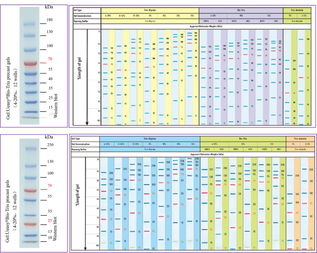
Color Prestained Protein Marker and Protein Migration Table
 

SKU
Name
Concentration
Wells
Ueasy®Bis-Tris precast gels(8%,12 wells)
8%
12 wells
Ueasy®Bis-Tris precast gels(8%,15 wells)
8%
15 wells
Ueasy®Bis-Tris precast gels(10%,12 wells)
10%
12 wells
Ueasy®Bis-Tris precast gels(10%,15 wells)
10%
15 wells
Ueasy®Bis-Tris precast gels(12%,12 wells)
12%
12 wells
Ueasy®Bis-Tris precast gels(12%,15 wells)
12%
15 wells
Ueasy®Bis-Tris precast gels(4-12%,12 wells)
4-12%
12 wells
Ueasy®Bis-Tris precast gels(4-12%,15 wells)
4-12%
15 wells
Ueasy®Bis-Tris precast gels(4-20%,12 wells)
4-20%
12 wells
Ueasy®Bis-Tris precast gels(4-20%,15 wells)
4-20%
15 wells
Ueasy®Tris-Glycine precast gels(8%,12 wells)
8%
12 wells
Ueasy®Tris-Glycine precast gels(8%,15 wells)
8%
15 wells
Ueasy®Tris-Glycine precast gels(10%,12 wells)
10%
12 wells
Ueasy®Tris-Glycine precast gels(10%,15 wells)
10%
15 wells
Ueasy®Tris-Glycine precast gels(12%,12 wells)
12%
12 wells
Ueasy®Tris-Glycine precast gels(12%,15 wells)
12%
15 wells
Ueasy®Tris-Glycine precast gels(4-12%,12 wells)
4-12%
12 wells
Ueasy®Tris-Glycine precast gels(4-12%,15 wells)
4-12%
15 wells
Ueasy®Tris-Glycine precast gels(4-20%,12 wells)
4-20%
12 wells
Ueasy®Tris-Glycine precast gels(4-20%,15 wells)
4-20%
15 wells

Note: This product is available in 12-well and 15-well formats. For custom well configurations (e.g., 10 wells, 20 wells, etc.), please contact our sales representative directly.

SKU
Product
Product Description
S-Blot®Power Pro
Universal power supply, Voltage: 5-600 V; Current: 1-1200 mA; Power: 1-500 W
W1506
V-Blot® Electrophoresis Unit Pro
Electrophoresis system, compatible with 4 gels; Glass plate size: 100×83 mm; Gel size: 83×73 mm
V-Blot®Electrophoresis Unit
Electrophoresis system, compatible with 2 gels; Glass plate size: 100×83 mm; Gel size: 83×73 mm
F-Blot®Mini Vertical Blot Pro
Western blot transfer system, compatible with 2 transfer cassettes; Transfer size: 110×90 mm; Built-in 2 cooling modules
T-Blot®Rapid Transfer Instrument
Fast transfer system, capable of transferring 4 gels simultaneously; Circulating cooling power supply directly cools the buffer for rapid temperature reduction
F-Blot® Mini Vertical Blot
Western blot transfer system, compatible with 2 transfer cassettes; Transfer size: 110×90 mm; Built-in 2 cooling modules
W2199
U-Blot® Rapid Sinking Loading Buffer(5×)
Instant loading buffer, no sample floating, fast thawing, odorless
U-Blot® Prestained Protein Marker(10-180kDa)
Sharp bands, ready-to-use, no extra handling required. Lowest molecular weight band at 10 kDa; multiple molecular weight ranges available
U-Blot® Prestained Protein Marker(10-250kDa)
Sharp bands, ready-to-use, no extra handling required. Lowest molecular weight band at 10 kDa; multiple molecular weight ranges available

Leave a Comment

Your email address will not be published. Required fields are marked *