LC3A Rabbit Monoclonal Antibody

LC3A Rabbit Monoclonal Antibody

Background:MAP1A and MAP1B are microtubule-associated proteins which mediate the physical interactions between microtubules and components of the cytoskeleton. MAP1A and MAP1B each consist of a heavy chain subunit and multiple light chain subunits. The protein encoded by this gene is one of the light chain subunits and can associate with either MAP1A or MAP1B. Two transcript variants encoding different isoforms have been found for this gene. The expression of variant 1 is suppressed in many tumor cell lines, suggesting that may be involved in carcinogenesis.

Category Item Description
Antibody & Materials Primary Antibody LC3A Rabbit mAb:WR4052
Antibody & Materials Nitrocellulose Membrane 0.22 Nitrocellulose Membrane:W3N21
Antibody & Materials Protein Marker pre-staining standard protein Marker(10-180kDa):W2100
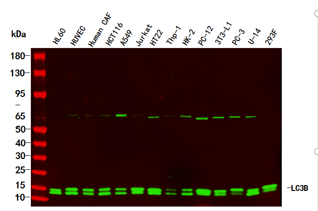
Lane Cell Type
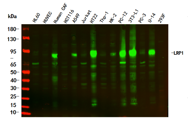
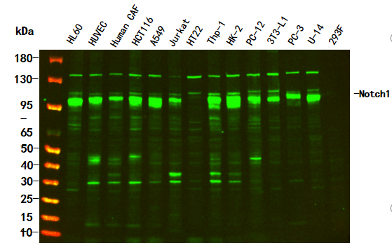
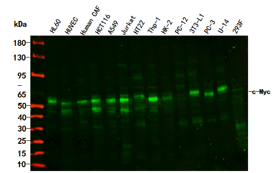
Lane1 HL60 – Human promyelocytic leukemia cell
Lane2 HUVEC – Human umbilical vein endothelial cell
Lane3 Human CAF – Human cancer-associated fibroblast
Lane4 HCT116 – Human colorectal carcinoma
Lane5 A549 – Human lung carcinoma
Lane6 Jurkat – Human T lymphocyte leukemia
Lane7 HT22 – Mouse hippocampal neuronal
Lane8 Thp-1 – Human monocytic leukemia
Lane9 HK-2 – Human proximal tubular epithelial
Lane10 PC-12 – Rat adrenal pheochromocytoma
Lane11 3T3-L1 – Mouse embryonic fibroblast cells
Lane12 PC-3 – Human prostate adenocarcinoma
Lane13 U-14 – Mouse cervical carcinoma
Lane14 293F – HEK293 derivative, adapted for suspension culture
Step Description
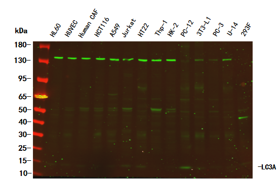
Electrophoresis Various whole cell lysates were separated by 4-20% SDS-PAGE
Primary Antibody Incubation The primary antibody was used at 4°C, over night with a 1:2500 dilution
Secondary Antibody Detection The Dylight 800-conjugated Goat anti-Rabbit antibody(Cat:WS0446) was used to detect the antibody
Type Size
Predicted band size 14 kDa
Observed band size 16 kDa

Elevate your autophagy and carcinogenesis research with the highly specific LC3A Rabbit Monoclonal Antibody. Validated across diverse cell lines and optimized for Western Blot, it’s the trusted tool for investigating LC3A-mediated microtubule-cytoskeleton interactions, autophagic pathways, and tumor cell expression dynamics. Accelerate discoveries in oncology and cell biology with this top-tier antibody today!
Leave a Comment

Your email address will not be published. Required fields are marked *